Glaucoma Runs in the Family

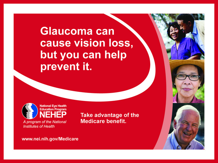
[heading centered=”yes” margin_bottom=”no”]Family History and Glaucoma[/heading]

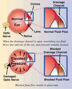
According to a study reported at the World Glaucoma Congress, the most efficient way to detect Primary Open-Angle Glaucoma (POAG), which is the most common form of glaucoma, is by knowing the medical history of close relatives with the disease. If you have a parent, sibling or child with POAG you have a greater than 25% chance of developing the disease. Others at risk include African Americans over 40, and anyone over 60, especially Mexican Americans. The earlier POAG is detected, the better the prognosis for retaining vision throughout one’s life. POAG occurs when the fluid that nourishes the eye cannot drain properly and the resulting increased pressure inside the eye damages the optic nerve. This is painless and not visually noticeable at first, but will lead to vision loss and blindness if left untreated. Increased pressure in the eye isn’t a sure sign of POAG, but it is a sign of increased risk for the disease. To read more about glaucoma click here.

Early diagnosis is of utmost importance. Dr. Griffith examines different parts of your eye to determine if you have or are at risk for developing POAG. Detection and diagnosis relies on tests for pressure within the eye (Tonometry), corneal thickness (Pachymetry) and observation of the optic nerve (Ophthalmoscopy), optovue
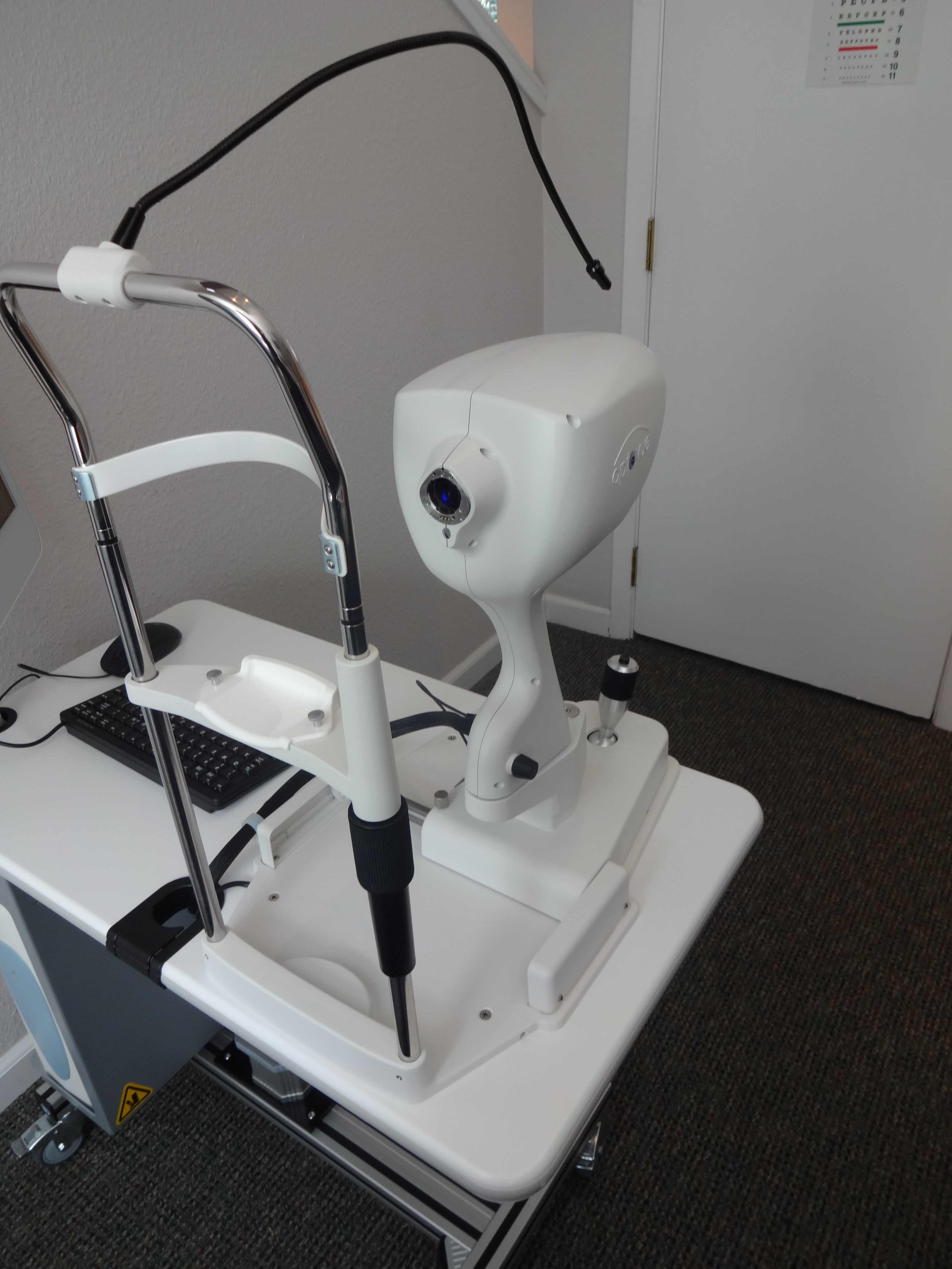
quantification of the retinal nerve fiber layer (Optical Coherence Tomography) and visual fields, including peripheral vision. Because of the increased risk among family members, a review of family history is also part of the screening. (Likewise, a positive diagnosis of POAG would be valuable information for close family members.)

Vision lost to glaucoma cannot be restored. The goal with treatment is to slow the disease progress and prevent further vision loss. The most common treatment is a prescription eye drop meant to reduce pressure in the eye. It is important for Dr. Griffith to be aware of other medications you are taking in order to find a compatible treatment for the glaucoma; fortunately there are usually several options. Additional treatment includes laser surgery or sometimes more traditional surgery; both physically alter and improve the drainage structure in the eye. In conclusion, regular eye examinations are vital to prevent unnecessary vision loss. If you are newly diagnosed with POAG or any form of glaucoma, Dr. Griffith can give you more detailed information.

What is Glaucoma?

Glaucoma is a group of eye diseases in which the pressure inside the eye may or may not be elevated. If untreated, vision loss or blindness may occur.

glaucoma prevention

January is Glaucoma Awareness Month
Unfortunately, the most common type of glaucoma – open angle, causes no symptoms. Vision doesn’t change, and there is no pain. However, as the disease progresses, a person with glaucoma may notice his or her side vision gradually failing. That is, objects in front may still be seen clearly, but objects to the side may be missed. As the disease worsens, the field of vision narrows and blindness results.
Studies have shown that early detection and treatment of glaucoma, before it causes major vision loss, is the best way to control the disease. So, if you fall into one of the high-risk groups for the disease, make sure to have your eyes examined annually. Individuals at high risk for glaucoma include African Americans over the age of 40, everyone over 60 and people with a family history of glaucoma.

There isn’t a single test to diagnose glaucoma. Drs. Griffith and Staton examine the appearance of the optic nerve, the intra-ocular pressure and visual field results. Further testing such as measurement of the corneal thickness (pachymetry), examination of the anterior chamber and measurement of the  integrity of the nerve fiber layer can be helpful in the diagnosis and treatment of glaucoma.

Schedule your appointment.

What is Macular Degeneration

Age Related Macular Degeneration (AMD) occurs in the macula in the back of the eye. The macula is part of the retina and contains the “cones,” a type of nerve cell responsible for clear vision and color vision. When AMD develops, the cones are disturbed and vision is significantly and negatively effected.
There are two types of AMD – wet and dry. The wet form is less common, but can progress quickly and profoundly. The symptoms are the same – blurred central vision, distorted central vision and missing areas of vision. A person with advanced AMD would not be able to distinguish facial features when looking straight ahead, but peripheral areas would still be visible.

Who gets AMD?

Many things contribute to the risk of developing AMD: age, race, family history, history of smoking, exposure to UV light, obesity, hypertension and a high fat diet. Some of these risks factors are controllable and prevention is recommended. Early AMD does not always have symptoms. Regular dilated eye exams can increase awareness and prevent devastating vision loss.

To read more about AMD symptoms, treatment and prevention click here.

January is Glaucoma Awareness Month

There are no symptoms for glaucoma. Vision stays normal and there is no pain. However, as the disease progresses, a person with glaucoma may notice a decrease in his or her side vision. Once vision is lost to glaucoma, it can’t be restored. That is why it is important to have regular eye examinations with pupil dilation.
Measurement of the “eye pressure” is just one factor in determining glaucoma. In fact, it is the damage to the optic nerve that confirms the diagnosis of glaucoma. The optic nerve is examined by dilating the pupil to view it with stereopsis (with depth). The dilation also allows better, clearer fundus (back of the eye) pictures to be taken for year-to-year comparison.

Glaucoma

Visual field testing measures the nerve fiber layer in the eye and can detect loss of important nerve tissue before optic nerve changes occur.

Studies have shown that early detection and treatment of glaucoma is the best way to control the disease. To learn more about glaucoma and how it is treated, click here.