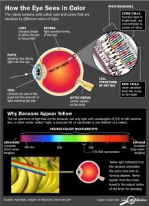
How the Eye Sees Color

Color VisionThe 6 to 7 million cones in the human retina are responsible for color vision. The cones are photoreceptors concentrated in the central zone of the retina called the macula. The center of the macula is called the fovea, and this tiny (0.3mm diameter) area contains the highest concentration of cones in the retina and is responsible for our most acute color vision.

Inherited forms of color blindness often are related to deficiencies in certain types of cones or outright absence of these cones. Color blindness is not a form of blindness at all, but a deficiency in the way we see color. With this vision problem, individuals have difficulty distinguishing certain colors, such as blue and yellow or red and green.

Color vision deficiency is an inherited condition that affects males more frequently than females. An estimated 8 percent of males and less than 1 percent of females have color vision problems.

Red-green color deficiency is the most common form of color deficiency. Red-green color blindness is caused by a common X-linked recessive gene. Your mother must be a carrier of the gene or be color deficient herself. Fathers with this inherited form of red-green color blindness pass the X-linked gene to their daughters but not their sons, because a son cannot receive X-linked genetic material from his father.

Any time a mother passes along this X-linked trait to her son, he will inherit the color vision deficiency and have trouble distinguishing reds and greens.

There is no cure for color blindness. But some coping strategies may help an individual function better in a color-oriented world. Most people are able to adapt to color vision deficiencies without too much trouble. But some professions should be avoided, such as graphic designer, ship pilots, interior decorator and other occupations that  require precise differentiation of colors and depend on accurate color perception.

If the color deficiency is identified early enough in life, the individual may be able to compensate by training for one of the many careers that are not as dependent on the ability to see in a full range of colors.

It is possible to develop color vision problems later in life. Sudden or gradual loss of color vision can indicate any number of underlying health problems, such as cataracts. Color blindness can occur when changes due to aging  damage retinal cells. An injury or damage to areas of the brain where vision processing takes place also can cause color vision changes. If you feel your perception of color is changing, schedule an eye exam.

At Westside Optometry we use a desaturated color vision test that takes a couple of minutes.

Color Vision Deficiency

Color vision deficiency is often called “color blindness” by mistake. Color vision deficiency describes a number of different problems people have with color vision. Abnormal color vision may vary from not being able to tell certain colors apart to not being able to identify any color.

Color vision deficiency affects an estimated 8% of males and fewer than 1% of females. Most color vision problems run in families and are inherited and present at birth.

A child inherits a color vision deficiency by receiving a faulty color vision gene from a parent. Abnormal color vision is found in a recessive gene on the X chromosome, therefore, color vision deficiency is a sex-linked condition. Because males only have one X chromosome, they are more likely to have color vision deficiencies than females.

Heredity does not cause all color vision problems. One common problem happens from the normal aging of the eye’s lens. The lens is clear at birth, but the aging process causes it to darken and yellow. Older adults may have problems identifying certain dark colors, particularly blues. Certain medications as well as inherited or acquired retinal and optic nerve disease may affect normal color vision.

At Westside Optometry, we test our young boys and some girls for color vision deficiency. Any child who is having difficulty in school should be checked for possible visual problems including color vision impairment. If there is a family history of color deficiency or your child is having problems identifying colors, let us know so he/she can be tested.

The specialized cells in the retina are called rods and cones. You use these cells for normal vision. Rods are useful for night vision and working in dim light. Cones are responsible for color vision. They work best in daylight. Three types of cone pigments are present in normal vision. These are sensitive to either blue, green or red colored objects. Together, they let you see a wide range of colors, from purple through red.

For normal color vision, all three cone pigments must work correctly. When a cone pigment is abnormal or missing, a type of color vision deficiency results. For example, the most common deficiency causes confusion between red and green colors. In rare cases, a person is born without any cones. These people are truly “color blind.” They see the world in shades of gray and suffer significant visual impairment. Most types of color vision deficiency are present at birth. There are also some types caused by aging, eye disease or injury.

Color Vision Testing

There are several ways to test color vision. Simpler tests involve colored figures (either shapes or numbers) placed against a busy, patterned background. A person with normal color vision can see the figures against the background. Those with color vision deficiencies cannot see the symbols.

At Westside Optometry we do a more specific test that requires placing colored disks in order, from one shade to another. People with different color deficiencies place the disks in varying order.

Unfortunately, there is no cure for hereditary color vision deficiency. Many people with color vision deficiency develop their own “system” or learn to identify colors by other means. Some people learn to tell colors apart by brightness and location. A stoplight, for example, is designed so the colors are always in the same order.


Color Vision Deficiency

Color vision deficiency is often called “color blindness” by mistake. Color vision deficiency describes a number of different problems people have with color vision. Abnormal color vision may vary from not being able to tell certain colors apart to not being able to identify any color.

Color vision deficiency affects an estimated 8% of males and fewer than 1% of females. Most color vision problems run in families and are inherited and present at birth.

A child inherits a color vision deficiency by receiving a faulty color vision gene from a parent. Abnormal color vision is found in a recessive gene on the X chromosome, therefore, color vision deficiency is a sex-linked condition. Because males only have one X chromosome, they are more likely to have color vision deficiencies than females.

Heredity does not cause all color vision problems. One common problem happens from the normal aging of the eye’s lens. The lens is clear at birth, but the aging process causes it to darken and yellow. Older adults may have problems identifying certain dark colors, particularly blues. Certain medications as well as inherited or acquired retinal and optic nerve disease may affect normal color vision.

At Westside Optometry, we test our young boys and some girls for color vision deficiency. Any child who is having difficulty in school should be checked for possible visual problems including color vision impairment. If there is a family history of color deficiency or your child is having problems identifying colors, let Dr. Griffith know so he/she can be tested.

The specialized cells in the retina are called rods and cones. You use these cells for normal vision. Rods are useful for night vision and working in dim light. Cones are responsible for color vision. They work best in daylight. Three types of cone pigments are present in normal vision. These are sensitive to either blue, green or red colored objects. Together, they let you see a wide range of colors, from purple through red.

For normal color vision, all three cone pigments must work correctly. When a cone pigment is abnormal or missing, a type of color vision deficiency results. For example, the most common deficiency causes confusion between red and green colors. In rare cases, a person is born without any cones. These people are truly “color blind.” They see the world in shades of gray and suffer significant visual impairment. Most types of color vision deficiency are present at birth. There are also some types caused by aging, eye disease or injury.

Color Vision Testing

There are several ways to test color vision. Simpler tests involve colored figures (either shapes or numbers) placed against a busy, patterned background. A person with normal color vision can see the figures against the background. Those with color vision deficiencies cannot see the symbols.

At Westside Optometry we do a more specific test that requires placing colored disks in order, from one shade to another. People with different color deficiencies place the disks in varying order.

Unfortunately, there is no cure for hereditary color vision deficiency. Many people with color vision deficiency develop their own “system” or learn to identify colors by other means. Some people learn to tell colors apart by brightness and location. A stoplight, for example, is designed so the colors are always in the same order.