How Does Glaucoma cause Vision Loss?
Doctors and researchers don’t know exactly how glaucoma damages the optic nerve. For many people, increased eye pressure seems to play an important role.
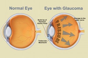
Your eye produces a watery fluid (aqueous humor), which goes into the eye and drains out. When your eye is healthy, the fluid drains through a mesh-like pathway and into the bloodstream. Aqueous fluid is produced by the ciliary body. It flows through the pupil and behind the clear cornea. Finally, it drains away through the trabecular meshwork.
 For some people, fluid can’t drain properly because of a faulty drainage system. Drainage that once worked well may gradually slow down as you get older, like a sink that becomes clogged and backs up with water. When there is no place for excess fluid to go, pressure inside the eye builds up.
This increased eye pressure may damage the optic nerve over time. Slowly, the nerve fibers that are essential for vision die.
For others, glaucoma damages the optic nerve without increased pressure. These people may be unusually sensitive even to normal levels of pressure. Their glaucoma may also be related to problems with blood flow in the eye. Doctors continue to study eye pressure and other possible causes of glaucoma.
Different people experience glaucoma differently. Usually, glaucoma affects side vision (peripheral vision) first. Late in the disease, glaucoma may cause “tunnel vision.” In this condition, the person can only see straight ahead. That’s why someone with glaucoma can have good central vision. However, even central vision can be seriously damaged.
How Does Dr. Griffith Check for Glaucoma?
There are three major signs that a person may have glaucoma:
- Optic nerve damage
- Increased eye pressure (elevated intraocular pressure).
- Vision loss(visual field loss)
These are some of the tests I will use to look for glaucoma:
Dilated Eye Exam
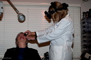
The doctor will place a few drops in your eye to open or dilate the pupil. This allows Dr. Griffith a clearer view to inspect the optic nerve at the back of the eye using Ophthalmoscopy.
Photography may be used to show the appearance of the optic nerve inside your eye. Photography also provides a record to help the doctor see changes from one year to the next.
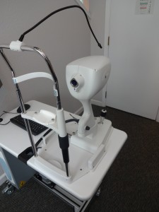
Tonometry measures pressure in the eye. Yellow colored drops are used to numb the eye. An instrument gently presses against the tear film on the surface of the eye. Pressure is shown as a number followed by the abbreviation “mm Hg.” This stands for “millimeters of mercury,” a standard measure for pressure. An average pressure is about 16 mm Hg. Still, a higher than average number doesn’t always mean you have glaucoma. Since the thickness of the cornea (the front window of the eye) may affect the pressure reading and the risk of glaucoma progression, Dr. Griffith may measure this as well. with a Pachymeter.
Perimetry evaluates your visual field. This tests your vision all around your field of view to see if any areas are have reduced sensitivity to light. It usually involves staring straight ahead at a target and trying to see lights that randomly appear in your periphery. This is generally done with a computerized system.
Optical Coherence Tomography or OCT scans the retina to measure the nerve fiber layer and ganglion cell complex.  Both are indicators of the health of the optic nerve and amount of healthy nerve tissue.
Gonioscopy allows a more accurate diagnosis of the type of glaucoma. After numbing the eye, the doctor gently places a special lens on the surface to examine the area in the front of the eye that drains fluid. Gonioscopy allows a more accurate diagnosis of the type of glaucoma.
How is Glaucoma treated?
Glaucoma can usually be treated and controlled using medicine, surgery a combination of these treatments. Medicated eye drops are typically the first step in treatment. The treatment will depend on the severity of the glaucoma and how the eye pressure responds to treatment.

3 thoughts to “Glaucoma”