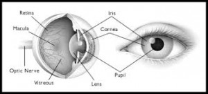
I’m going to give you a tour of the human eye. When I do cow eye dissections for children, I identify structures from the front of the eye to the back of the eye, the same way light goes through the eye.
First and most visible are the eyelids and eyelashes. The eyelashes work like a screen to deflect debris and dust from our eyes. The eyelids provide a barrier to foreign objects. The eyelids also work like windshield wipers to smooth tears across the eye and move debris off of the surface.
The cornea is the clear part of the eye and most sensitive to pain. It is important that the cornea maintain it’s clarity for us to see clearly. If it becomes scratched or scarred, our vision becomes obscured. The cornea is the window to the eye.
The conjunctiva is the white of the eye and the protective covering in the front of the eye. The sclera is the protective layer that covers the whole eye.
Directly behind the cornea is a fluid filled space called the anterior chamber. It is filled with aqueous humor, a watery solution.
The colored part of the eye or the iris is next. It can be different colors depending on the amount of pigmentation. The iris constricts or dilates to decrease or increase the size of the pupil. The pupil is actually a hole or aperture.
The crystalline lens is behind the iris. It adjusts to focus the light before it reaches the retina.
Filling the eyeball between the lens and the retina is a gel-like substance called the vitreous humor. This is where “floaters” are located.
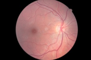
The retina is composed of an intricate nerve layer. It has a super sensitive area called the macula. Most of the cones (photoreceptors) are located in this small area to provide the clearest vision and color vision. All the nerves come together at the optic nerve head. From the optic nerve head in the eye, impulses travel through the optic nerve to the brain.


3 thoughts to “Eye Anatomy”