Pinguecula and Pterygium
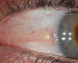
There are several types of bumps that can occur in the eye. PINGUECULA are the most common and occur on the white of the eye. They may be yellow, pink or white and are usually located on the white part of the eye, or conjunctiva, in the space between the eyelids, almost always on the side closes to the nose.
A pinguecula is a benign growth caused by the degeneration of the conjunctiva’s collagen fibers. Thicker yellow tissue and in some cases calcified deposits, eventually replace the original fibers. The pinguecula can become irritated, especially if exposed to dust, wind and sun. The exact cause or causes of this disorder are debated, but it occurs more frequently in people who live in sunny and windy climates and people whose jobs expose them to ultraviolet (UV) light.
Most people with pinguecula do not require treatment unless their symptoms are severe. Lubricating eye drops are normally recommended to relieve irritation and the feeling that there is something that doesn’t belong in the eye.
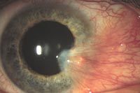
A less common type of eye bump is a PTERYGIUM (don’t pronounce the “p”). A pterygium is a triangular shaped growth on the white part of the eye that also extends onto the clear part of the eye, or cornea. A pterygium contains blood vessels and can be of greater cosmetic concern than the pinguecula. In extreme cases, pterygia may grow far enough onto the cornea to interfere with vision. A pterygium can result from a pinguecula that continues to grow. Most bumps grow slowly and almost never cause significant damage. Diagnosis should be made by an eye doctor to rule out other more serious disorders.
Most people with a pterygium are asymptomatic but others can experience a significant feeling of having something irritating in the eye. Because a pterygium can distort the cornea, some people develop astigmatism from the condition; if the astigmatism is significant, reduced vision can result.
Usually no treatment is indicated. Artificial tears can be used to relieve any dry sensation and reduce redness. Surgery to remove the pterygium is considered when the effect on the cornea reduces vision or when the growth is causing excessive and recurrent discomfort or inflammation. Surgical removal can also be for cosmetic reasons, however it is important to realize that healing from this type of surgery, although usually painless, takes many weeks, and there is a high rate of growth recurrence, as high as 50-60%. Therefore, surgery is usually not recommended unless discomfort is significant or vision is affected.
There is nothing that has been clearly shown to prevent these disorders, or to prevent a pinguecula from progressing to a pterygium. However, the presence of these bumps has been linked to exposure to UV light. For that reason, UV exposure should be minimized whenever possible by wearing good sunglasses.
Subconjunctival Hemorrhage

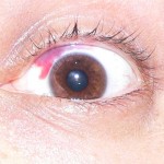
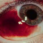
Many of us have had a blood red spot on the white of the eye at one time or another. Usually there was no injury or discomfort. Many people wake-up with the red spot and may not even be aware of it until a family member or co-worker asks about it. Subconjunctival hemorrhages look worse than they are and do not need treatment.
The conjunctiva is a thin membrane that covers the surface of the inner eyelid and the white part of the eyeball. The conjunctiva contains many small, fragile blood vessels that are easily ruptured or broken. Subconjunctival hemorrhage occurs when a small blood vessel in the conjunctiva breaks and bleeds. It may occur spontaneously or from heavy lifting, coughing or vomiting. In some cases, it may develop following eye surgery or trauma. Subconjunctival hemorrhage tends to be more common among those with diabetes and hypertension.
Certain medications can make the bleeding worse, including: Coumadin, Aspirin, Plavix, St. John’s Wort and Ginkgo.
While it may look frightening, a subconjunctival hemorrhage is essentially harmless. The blood from the broken conjunctival vessel becomes trapped in the space underneath the clear conjunctival tissue. The blood naturally absorbs within one to three weeks and may turn greenish or yellow during this time.
A subconjunctival hemorrhage does not affect vision or cause pain, and treatment is usually not required. Exceptions to that are when bleeding: is a result of trauma or injury; lasts more than 10 days; starts again.
And of course, if you are unsure call our office. 
Family History and Glaucoma
According to a study reported at the World Glaucoma Congress, the most efficient way to detect Primary Open-Angle Glaucoma (POAG), which is the most common form of glaucoma, is by knowing the medical history of close relatives with the disease. If you have a parent, sibling or child with POAG you have a greater than 25% chance of developing the disease. Others at risk include African Americans over 40, and anyone over 60, especially Mexican Americans. The earlier POAG is detected, the better the prognosis for retaining vision throughout one’s life. POAG occurs when the fluid that nourishes the eye cannot drain properly and the resulting increased pressure inside the eye damages the optic nerve. This is painless and not visually noticeable at first, but will lead to vision loss and blindness if left untreated. Increased pressure in the eye isn’t a sure sign of POAG, but it is a sign of increased risk for the disease. To read more about glaucoma click here.
Early diagnosis is of utmost importance. Dr. Griffith examines different parts of your eye to determine if you have or are at risk for developing POAG. Detection and diagnosis relies on tests for pressure within the eye (Tonometry), corneal thickness (Pachymetry) and observation of the optic nerve (Ophthalmoscopy) and visual fields, including peripheral vision. Because of the increased risk among family members, a review of family history is also part of the screening. (Likewise, a positive diagnosis of POAG would be valuable information for close family members.)
Vision lost to glaucoma cannot be restored. The goal with treatment is to slow the disease progress and prevent further vision loss. The most common treatment is a prescription eye drop or pill meant to reduce pressure in the eye. It is important for Dr. Griffith to be aware of other medications you are taking in order to find a compatible treatment for the glaucoma; fortunately there are usually several options. Additional treatment includes laser surgery or sometimes more traditional surgery; both physically alter and improve the drainage structure in the eye. In conclusion, regular eye examinations are a vital to prevent unnecessary vision loss. If you are newly diagnosed with POAG or any form of glaucoma, Dr. Griffith can give you much more detailed information.


One thought to “Eye Health”