Blog

Styes

Close up right upper eye lid abscess "stye or hordeolum"

There are 2 types of red and painful eyelid lumps:
STYES are at the base of the eyelashes in the tiny opening of the oil-producing glands. The oily secretions from these glands are an essential component of the tears and help keep the eyes lubricated. If the openings become clogged, bacteria grows and a localized infection called a STYE results.
CHALAZIA look similar to a stye but affect the meibomian glands which are located deeper in the eyelids. A chalazion usually does not contain an active bacterial infection. It may initially be red and swollen but within a few days it changes to a painless, slow growing lump within the eyelid.

Treatment

Styes are usually treated with hot, moist compresses. Since the stye is caused by bacteria, usually from our hands or make-up, I recommend replacing your makeup and of course not rubbing your eyes.

Chalazia are also treated with hot moist compresses but more aggressively, 10 minutes four times a day. This may soften the debris trapped in the duct and promote drainage. Chalazia are often associated with blepharitis or eyelid inflammation and preventative steps are recommended.

Keep Your Vision Healthy

Clear View

Did you know your eyes are windows to your general health? Many systemic conditions can be detected with a dilated eye examination. Looking into a dilated eye, I can see a view of the blood vessels and assess vascular health. Your eyes can tell a lot about your visual health and overall wellness.
Besides helping you see better, annual eye exams can aid in detection of serious eye conditions, like glaucoma and cataracts and health conditions like diabetes and high blood pressure. This is important since you won’t always notice the symptoms yourself – and some of these diseases cause irreversible damage.

If it’s been at least a year since your last eye examination, call the office and schedule an appointment. (707)762-8643. Schedule online.

How to Clean Your Glasses


The following tips are intended to provide you clean eyeglass lenses and frames without the risk of scratching the lenses or damaging the frame.

  1. Wash and dry your hands. Before cleaning your eyeglasses, make sure your hands are free from dirt, grime and lotion. Use lotion-free soap or dishwashing liquid and a clean, lint-free towel to clean your hands clean eyeglasses
  2. Rinse your glasses under a gentle stream of lukewarm tap water. This will remove dust and other debris, which can help avoid scratching your lenses when you are cleaning them. Avoid hot water, which can damage some lens coatings.
  3. Apply a small drop of lotion-free dishwashing liquid to each lens. Most dishwashing liquids are very concentrated, so use a tiny amount. Or apply a drop to your fingertip instead.
  4. Gently rub both sides of the lenses and all parts of the frame for a few seconds. Make sure you clean every part, including the nose pads and ends of the temples that rest behind your ears. And be sure to clean the area where the edge of the lenses meet the frame where dust, debris and skin oils can accumulate.
  5. Rinse both sides of the lenses and the frame thoroughly. Failing to remove all traces of the soap will cause the lenses to be smeared when you dry them.
  6. Use a clean lint-free towel to dry the lenses and frame. Avoid rough fabrics and paper products which can scratch the lenses and the finish on the frame.

Are Rigid Gas Permeable Contacts Old-Fashioned?

There are many contact lens choices. At Westside Optometry we will prescribe the best lens for you, your eyes and your lifestyle.

RGP, Soft, Hybrid, Scleral

The majority of contact lenses prescribed are soft lenses. Within this large category of lens are daily disposables, weekly and monthly replacement. Functional options include multifocals for presbyopia, torics for astigmatism and spherical lenses for hyperopia and myopia.
What happens if there is “a lot” of astigmatism, or astigmatism AND presbyopia or high myopia or high hyperopia? And what if the cornea has been altered by disease or refractive surgery?
Soft contact lenses can’t correct many of these conditions. That’s why there are other contact lens options. Gas Permeable (GP) contact lenses correct astigmatism, have stable crisp optics and are individually designed and manufactured. GP lenses provide more oxygen to the cornea than soft lenses. They last longer and are more durable providing a cost effective solution. Gas Permeables are available in high powers, multi-focals and special designs for irregular corneas.
Hybrids are a blend of soft and gas permeable lens designs. The center of the hybrid lens is gas permeable material and the perimeter is a silicone hydrogel material. This provides crisp optics of the GP and the comfort of a soft lens. The hybrid lenses are available in multifocals and special designs for post-LASIK and irregular corneas.

The majority of contact lens wearers use a soft contact lens, but gas permeable contacts  have many benefits and applications. Gas permeable contact lenses are not old-fashioned,  they have stood the test of time.

What is Macular Degeneration?

Macular degeneration is a retinal disease which can lead to legal blindness. More specifically, it affects the macula, the portion of the retina important for detailed vision like reading.

There are two forms of macular degeneration: wet and dry. Dry macular degeneration is usually slower in destruction of the macula, causing gradual to little loss of central vision. The wet form is more aggressive and destroys the central vision faster. The wet form is associated with the presence of abnormal blood vessels growing somewhere within the layers of the retina. These abnormal blood vessels can leak and bleed.

ARMD wet before treatment

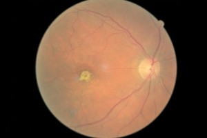
Dry macular degeneration causes a slower loss of tissue. While there is treatment for the wet form, there is no approved treatment for the dry form.

Drusen (800x533)

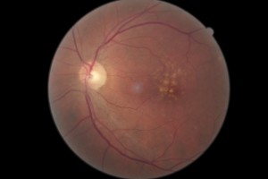
Symptoms of Macular Degeneration include blurry central vision and/or distortion. In some cases, there may be blind spots within the central vision. The vision loss in both types is progressive. It is the rate at which vision loss develops which differentiates the wet form from the dry. Remember, wet macular degeneration causes faster and more devastating loss of vision compared to the dry form.

Treatment for Macular Degeneration

There is no treatment for dry macular degeneration. While AREDS (Age-Related Eye Disease Study) vitamins may be indicated for a small subset of macular degeneration patients, it does not improve the condition or vision. The supplements are primarily preventative. There are different formulations of “eye vitamins,” but the contents of the supplement used for the study were 500 milligrams of vitamin C; 400 International Units of vitamin E; 15 milligrams of beta-carotene (often labeled as equivalent to 25,000 International Units of vitamin A); 80 milligrams of zinc as zinc oxide; and two milligrams of copper as cupric oxide. Copper was added to the AREDS formulations containing zinc to prevent copper deficiency anemia, a condition associated with high levels of zinc intake.

Other preventative measures for both types of macular degeneration include not smoking, regular exercise and a healthy diet.

The mainstay of treatment for wet macular degeneration involves injections into the eye with anti-VEGF (Vascular Endothelial Growth Factor) medications.

These medications (Lucentis, Avastin, Eylea) attach to the abnormal blood vessels, preventing additional leakage and bleeding. Often there can be improvement in vision.

Early treatment is key to maintaining vision. Regular dilated eye examinations are advised.

Leafy Green Vegetables and Glaucoma

New research indicates that leafy greens may be healthier than we thought. Eating vegetables like kale, spinach and collard greens may not cure glaucoma but they may be protective against developing primary open angle glaucoma(POAG). The research revealed that people who ate a lot of leafy greens had a 40 to 50 percent lower risk of acquiring POAG.


The reason these super foods offer such great protection is related to the dietary nitrate they contain. It’s thought that glaucoma impairs blood flow to the optic nerve. Nitric oxide helps regulate this flow. Since leafy greens contain high levels of nitrate, the precursor to nitric oxide, consuming them likely keeps things flowing more smoothly.

chard A significant amount of other new research is currently aimed at developing therapeutics that treat glaucoma by way of nitric oxide. In fact, the FDA is reviewing at least one new medication that donated nitric oxide. But thanks to this latest study, far fewer people will need it if they load up on their leafy green vegetables before any glaucomatous damage is done.

So how much roughage do you need to eat to protect yourself from glaucoma? In this study, those who consumed the most leafy greens, averaged about 1.5 servings per day, which equates to about one and a half cups. Leafy greens include kale, spinach, dandelion greens, romaine lettuce, radicchio, leaf lettuce, arugula, Swiss chard, rapini (broccoli rabe) and collard greens.

According to the book, EyeFoods, by Drs. Campagna and Pelletier, the leafy greens are the rockstars of eye nutrients. They include lutein, zeaxanthin, beta-carotene, vitamin E, vitamin C, zinc and fiber. If you’re not accustomed to the tastes and textures of leafy green vegetables, start slowly. Begin with some lettuce leaves or spinach. Add a few leaves of kale or arugula to a salad or soup. Work your way toward at least a serving per day.

Bumps on the White of the Eye

There are two types of benign growths that can appear on the white of the eye (conjunctiva).
eye bumpThe first type is called a Pinguecula. A pinguecula is a benign growth caused by the degeneration of the conjunctiva’s collagen fibers. Thicker yellow tissue and in some cases calcified deposits, eventually replace the original fibers. It often appears as a thickened, discolored spot between the colored part of the eye and the nose. It is more common in individuals who spend a lot of time outdoors with continued exposure to the ultraviolet light or those who live in sunny, windy equatorial climates. The affected eye might become red and irritated. No treatment is necessary unless discomfort is severe. Prevention is highly recommended and that means sunglasses when outdoors. Artificial tears can help with irritation.

The second type of benign bump on the eye can actually be more problematic. A Pterygium is an elevated, triangular, non-cancerous Pterygiumgrowth that is also usually on the nasal side of the eyeball. A pterygium contains blood vessels and can be of greater cosmetic concern than the pinguecula. The difference between a pterygium and a pinguecula is the tissue they invade. The pinguecula is limited to the conjunctiva and the pterygium grows onto the cornea (clear part of the eye). A small pterygium is treated with artificial tears for comfort and sunglasses for prevention. Sometimes surgery is needed if the vision is affected.

Genetics can play a role in the possibility of having either of these bumps. Wearing protection from UV is your best chance of preventing or controlling pinguecula and pterygia.


Facts About Eye Color

Girl colorful and natural rainbow eye on white background

Genes and pigment concentration are responsible for the color of a person’s eyes. High concentrations of melanin result in darker eye colors. Less melanin results in lighter eye colors. A special pigment called “lipochrome,” is responsible for some eye colors, like green.

By far the most common eye color is brown. In rank order the most common colors are:

  • 55% Brown
  • 8% Blue
  • 5-8% Hazel
  • 5% Amber
  • 5% Grey
  • 2% Green

BROWN 

The original eye color was brown and is evidence of a high concentration of a pigment called melanin in the iris stroma (the spongy part).

BLUE

Most caucasian babies are born with blue eyes; this color typically changes into the “adult” color after 6 months based on melanin concentration and exposure to the sun. A genetic mutation allows some humans to keep their blue eyes as adults. Blue eyes are the result of the back layer of the eye being dark and the middle spongy layer called the stoma lacking pigment.

HAZEL

Hazel eyes are the least understood and are thought to have originated in Europe. Some scientists believe that hazel is a mix of green and brown. This eye color is most common in the US and Europe and almost unheard of in Asia and Africa.

GREEN

Green eyes are one of the rarest. It is thought that this eye color originated in Europe, specifically in areas around Germany or Hungary. However, they were also around in Siberia during the Bronze Age. The green color is caused by a high concentration of the yellow pigment lipochrome – the same pigment responsible for Amber.

AMBER 

Amber eyes are mostly found in African and Asian countries where it is assumed the eye color originated. Noted for a yellow colored pigment, lipochrome which is responsible for the deep rich copper or golden amber color.

GREY

Silver or gray eyes are the rarest of all. They are thought to have origianted in Europe along with blue eyes. They are the result of low concentration of melanin in the eye.

RED

Red eyes are a result of a rare genetic mutation associated with albinism. There is little to no melanin in the iris which means people with red eye can’t block light from entering the iris.

Glaucoma Runs in the Family

[heading centered=”yes” margin_bottom=”no”]Family History and Glaucoma[/heading]

According to a study reported at the World Glaucoma Congress, the most efficient way to detect Primary Open-Angle Glaucoma (POAG), which is the most common form of glaucoma, is by knowing the medical history of close relatives with the disease. If you have a parent, sibling or child with POAG you have a greater than 25% chance of developing the disease. Others at risk include African Americans over 40, and anyone over 60, especially Mexican Americans. The earlier POAG is detected, the better the prognosis for retaining vision throughout one’s life. POAG occurs when the fluid that nourishes the eye cannot drain properly and the resulting increased pressure inside the eye damages the optic nerve. This is painless and not visually noticeable at first, but will lead to vision loss and blindness if left untreated. Increased pressure in the eye isn’t a sure sign of POAG, but it is a sign of increased risk for the disease. To read more about glaucoma click here.

Early diagnosis is of utmost importance. Dr. Griffith examines different parts of your eye to determine if you have or are at risk for developing POAG. Detection and diagnosis relies on tests for pressure within the eye (Tonometry), corneal thickness (Pachymetry) and observation of the optic nerve (Ophthalmoscopy), optovuequantification of the retinal nerve fiber layer (Optical Coherence Tomography) and visual fields, including peripheral vision. Because of the increased risk among family members, a review of family history is also part of the screening. (Likewise, a positive diagnosis of POAG would be valuable information for close family members.)

Vision lost to glaucoma cannot be restored. The goal with treatment is to slow the disease progress and prevent further vision loss. The most common treatment is a prescription eye drop meant to reduce pressure in the eye. It is important for Dr. Griffith to be aware of other medications you are taking in order to find a compatible treatment for the glaucoma; fortunately there are usually several options. Additional treatment includes laser surgery or sometimes more traditional surgery; both physically alter and improve the drainage structure in the eye. In conclusion, regular eye examinations are vital to prevent unnecessary vision loss. If you are newly diagnosed with POAG or any form of glaucoma, Dr. Griffith can give you more detailed information.

Red Spot on the Eye

Many of us have had a blood red spot on the white of the eye at one time or another. Usually there was no injury or discomfort. Many people wake-up with the red spot and may not even be aware of it until a family member or co-worker points it out. Subconjunctival hemorrhages look worse than they are and do not need treatment.

moderate blood on eye The conjunctiva is a thin membrane that covers the inner surface of the eyelid and the white part of the eyeball. The conjunctiva contains many small, fragile blood vessels that are easily ruptured or broken. Subconjunctival hemorrhage occurs when a small blood vessel in the conjunctiva breaks and bleeds. It may occur spontaneously or from heavy lifting, coughing or vomiting. In some cases, it may develop following eye surgery or trauma. Subconjunctival hemorrhage tends to be more common among those with diabetes and hypertension.

Certain medications can make the bleeding worse, including: Coumadin, Aspirin, Plavix, St. John’s Wort and Ginkgo.

While it may look frightening, a subconjunctival hemorrhage is essentially harmless. The blood from the broken conjunctival vessel becomes trapped in the space underneath the clear conjunctival tissue. The blood naturally absorbs within one to three weeks and may turn greenish or yellow during this time.

A subconjunctival hemorrhage does not affect vision or cause pain, and treatment is usually not required. Exceptions are when the bleeding is a result of trauma or injury, the redness lasts more than 10 days or when it recurs again.

And of course, if you are unsure call our office. (707)762-8643