January is Glaucoma Awareness Month

There are no symptoms for glaucoma. Vision stays normal and there is no pain. However, as the disease progresses, a person with glaucoma may notice a decrease in his or her side vision. Once vision is lost to glaucoma, it can’t be restored. That is why it is important to have regular eye examinations with pupil dilation.
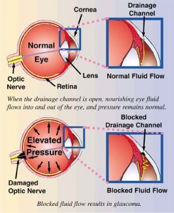
Measurement of the “eye pressure” is just one factor in determining glaucoma. In fact, it is the damage to the optic nerve that confirms the diagnosis of glaucoma. The optic nerve is examined by dilating the pupil to view it with stereopsis (with depth). The dilation also allows better, clearer fundus (back of the eye) pictures to be taken for year-to-year comparison.

Glaucoma

Visual field testing measures the nerve fiber layer in the eye and can detect loss of important nerve tissue before optic nerve changes occur.

Studies have shown that early detection and treatment of glaucoma is the best way to control the disease. To learn more about glaucoma and how it is treated, click here.


Westside Optometry Expands Services

I’m going to crow a little here. The California State Board of Optometry notified me that I’m officially licensed to provide glaucoma care. This is an endeavor I started 10 years ago with Dr. Karen Griffithcourse work at UC Berkeley. I put it on hold because the state requirements were insurmountable. Laws changed and last January I made the decision to finish what I started with more coursework through UC Berkeley. After hours of online cases and tests (how things changed in 10 years) and two days of grand rounds at the  School of Optometry I had completed all the education and patient cases.
Most of my patients won’t experience any difference in their care. But for those who I’ve been observing more closely for suspicious glaucoma findings, I won’t have to refer you to someone else as I can now treat the disease.


Maintaining Clear Vision

Age-related eye diseases such as macular degeneration and cataracts commonly cause impaired vision and blindness in older adults. But lifestyle changes, including good nutrition, could help delay or prevent certain eye problems.
Besides adopting a healthy diet, you also can protect your eyes by avoiding intense ultraviolet (UV) light, quitting smoking and getting regular checkups that may help detect chronic diseases contributing to eye problems. Diabetes, for example, increases your risk for age-related eye diseases and may cause diabetic retinopathy.
Regular eye exams, too, are essential for maintaining eye health as you grow older. If eye problems and chronic diseases are detected early enough, appropriate treatment may prevent permanent vision loss.

What is Glaucoma?

January is glaucoma awareness month. Glaucoma is often called “the sneak thief of sight.” That’s because people usually do not notice any signs of the disease until they have already lost significant vision. Once lost, vision can’t be restored. More than 2.7 million Americans age 40 and older have open angle glaucoma, the most common form of glaucoma. At least half don’t even know they have it.

Glaucoma is an eye disease that causes loss of sight by damaging a part of the eye called the optic nerve. This nerve sends information from your eyes to your brain. When glaucoma damages your optic nerve, you begin to lose patches of vision, usually side vision (peripheral vision). Over time, glaucoma may also damage the central vision. You may not notice a loss of side vision until you have lost a great deal of your sight. When checking for glaucoma, I measure the intra-ocular pressure, dilate the pupil to look for damage to the optic nerve and test peripheral vision. To learn more about how I identify glaucoma and how it is treated, click here

Who is at risk for glaucoma?

Below are the risk factors for glaucoma:

Age – The older you are, the greater you are at risk (especially if you are over 60 years old). African Americans are at a greater risk at a younger age starting at age 40 and older.

Race – African Americans age 40 and over are 4-5 times more likely to have glaucoma than others. Hispanics are also at increased risk for glaucoma as they age. Those of Asian and Native American descent are at increased risk for angle closure glaucoma.

Family history – If you have a parent, brother or sister with glaucoma, you are more likely to get glaucoma. If you have glaucoma, inform your family members to get complete eye exams.

Medical history – You are at risk if you have a history of high pressure in your eyes, previous eye injury, long term steroid use, or nearsightedness.

There are no symptoms for glaucoma until irreversible vision loss occurs. Have your eyes check.

High Blood Pressure and the Eyes

High Blood Pressure (HBP) is a serious condition that can lead to coronary heart disease, heart failure, stroke, kidney failure and vision loss. “Blood Pressure” is the force of blood pushing against the walls of the arteries as the heart pumps blood. If this pressure rises and stays high over time, it can damage the body in many ways.

About 1 in 3 adults in the United States has HBP. The condition itself usually has no signs or symptoms. You can have it for years without knowing it. During this time, the HBP can damage your heart, blood vessels, kidneys and eyes.

Blood Pressure tends to rise with age. Sometimes medication can control it. Following a healthy lifestyle helps people delay, prevent or control the BP.

Hypertension can cause damage to the blood vessels in the retina. This picture of the retina shows an eye with hypertensive retinopathy. The arrows point to arteries and veins crossing each other. In a healthy eye the vessels run parallel. The crossings are dangerous because one of the vessels can be occluded. If an artery is blocked, blood will not flow to the retina beyond the occlusion and the retina will die. This causes a permanent blind spot. If a vein is occluded there will be bleeding which may cause a chain of negative events including loss of vision, abnormal vessel growth and glaucoma.

Hypertensive retinopathy typically won’t have any symptoms. It is found during a dilated eye exam. The earlier it is detected the sooner the blood pressure can be controlled preventing vision loss, stroke and death.

The best way to prevent hypertensive retinopathy is by keeping blood pressure controlled by diet, exercise and taking medications as prescribed.

Save Your Vision Month

March is Save your Vision Month

It is important to be proactive and protect your eyesight and vision for the future. 

For adults in their 20s and 30s, the eyes are generally healthy as are the other organs in the body. Start protecting your vision now.

 Eat right! Include lots of fruits and vegetables in your diet
 Quit smoking
 Wear sunglasses to protect against harmful ultraviolet radiation.

For adults in their 40s and 50s, you are probably noticing some significant changes in vision, especially close-up. Menopause can increase dry eye symptoms for women.

 Ask family members about eye diseases. Many conditions run in the family
 Invest in accurate and quality reading glasses
 Your general health can have profound effects on your eyes. Be prepared to discuss medications and health conditions with your optometrist

After 60 it is crucial to maintain regular eye examinations. The incidence of many eye diseases increases. Many of these conditions like glaucoma, have no symptoms.

 Schedule annual exams or as recommended.
 Report all changes in general health to the eye doctor. Don’t accept declining vision as just a part of aging.

Visual Field Testing

Visual Field Testing

A perimeter is used to test your visual field. Your visual field includes both your central and side vision. During this test, while you are seated comfortably, you will be asked to look straight ahead at a central fixation target. You will be instructed to press a button when you become aware of a small shimmering light anywhere within your peripheral field. It’s important that you keep your eyes focused on the central target throughout this examination so that an accurate reading of your visual field is obtained.

This test can detect vision loss, even if the loss is far to the side or quite subtle. Even a large and profound vision loss in one eye’s visual field can go unnoticed by a patient because the visual field of the opposite eye overlaps it to a great extent. One eye simply “fill in” the area of vision loss present in the opposite eye. This is why the side-vision test is always administered to each eye separately.

The nerve pathways from the retina to the part of the brain where vision is interpreted are very uniform from person to person. Because of this uniformity, visual field analysis has a great deal of diagnostic value. Glaucomatous visual field loss usually follows a particular pattern. Other diagnostic patterns of visual field loss can indicate retinal detachment and neurological diseases, adding significantly to the value of this test. The nerve pathways for vision are long and cross over many brain structures on their route from the eye to the very back of the brain (visual cortex). Neurologists make great use of visual fields to help them determine where in the brain a tumor or other pathology is located.

Family History and Glaucoma

Family History and Glaucoma

Eye examinationAccording to a study reported at the World Glaucoma Congress, the most efficient way to detect Primary Open-Angle Glaucoma (POAG), which is the most common form of glaucoma, is by knowing the medical history of close relatives with the disease. If you have a parent, sibling or child with POAG you have a greater than 25% chance of developing the disease. Others at risk include African Americans over 40, and anyone over 60, especially Mexican Americans. The earlier POAG is detected, the better the prognosis for retaining vision throughout one’s life. POAG occurs when the fluid that nourishes the eye cannot drain properly and the resulting increased pressure inside the eye damages the optic nerve. This is painless and not visually noticeable at first, but will lead to vision loss and blindness if left untreated. Increased pressure in the eye isn’t a sure sign of POAG, but it is a sign of increased risk for the disease. 

Early diagnosis is of utmost importance. Dr. Griffith examines different parts of your eye to determine if you have or are at risk for developing POAG. Detection and diagnosis relies on tests for pressure within the eye (Tonometry), corneal thickness (Pachymetry) and observation of the optic nerve (Ophthalmoscopy) and visual fields, including peripheral vision. Because of the increased risk among family members, a review of family history is also part of the screening. (Likewise, a positive diagnosis of POAG would be valuable information for close family members.)

Vision lost to glaucoma cannot be restored. The goal with treatment is to slow the disease progress and prevent further vision loss. The most common treatment is a prescription eye drop or pill meant to reduce pressure in the eye. It is important for Dr. Griffith to be aware of other medications you are taking in order to fi nd a compatible treatment for the glaucoma; fortunately there are usually several options. Additional treatment includes laser surgery or sometimes more traditional surgery; both physically alter and improve the drainage structure in the eye. In conclusion, regular eye examinations are a certain age is vital to prevent unnecessary vision loss. If you are newly diagnosed with POAG or any form of glaucoma, Dr. Griffi th can give you much more detailed information.