How to Prevent Eye Complications due to Diabetes

There are steps you can take to avoid eye problems due to diabetes.

First and most important, keep your blood sugar levels under tight control. In the Diabetes Control and Complications Trial, people on standard diabetes treatment got retinopathy four times as often as people who kept their blood sugar levels close to normal. In people who already have retinopathy, the condition progressed in the tight-control group only half as often.

These impressive results show that you have a lot of control over what happens to your eyes. Also, high blood sugar levels may make your vision temporarily blurry.

Second, keep blood pressure under control. High blood pressure can make eye problems worse.

Third, quit smoking.

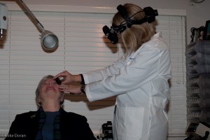
Fourth, see your optometrist at least once a year for a dilated eye exam. Having your regular doctor look at your eyes is not enough.

Fifth, see your optometrist if:

  • your vision becomes blurry
  • you have trouble reading signs or books
  • you see double
  • one or both of your eyes hurt
  • your eyes get red and stay that way
  • you feel pressure in your eye
  • you see spots or floaters
  • straight lines do not look straight
  • you can’t see things to the side as you used to

Don’t procrastinate. If you have diabetes and haven’t had a dilated eye exam in the last 12 months, schedule an eye exam now.

For more information, check out the American Diabetes Association.


Diabetes Myths

November is Diabetes Awareness Month.

To begin the  month of diabetes awareness, let’s start by dispelling some myths:

Myth: Diabetes is not that serious of a disease.

Fact: Diabetes causes more deaths a year than breast cancer and AIDS combined. Two out of three people with diabetes die from heart disease or stroke.

Myth: If you are overweight or obese, you will eventually develop type 2 diabetes.

Fact: Being overweight is a risk factor for developing this disease, but other risk factors such as family history, ethnicity and age also play a role. Unfortunately, too many people disregard the other risk factors for diabetes and think that weight is the only risk factor for type 2 diabetes. Most overweight people never develop type 2 diabetes, and many people with type 2 diabetes are at a normal weight or only moderately overweight.

Myth: Eating too much sugar causes diabetes.

Fact: The answer is not so simple. Type 1 diabetes is caused by genetics and unknown factors that trigger the onset of the disease; type 2 diabetes is caused by genetics and lifestyle factors.

Being overweight does increase your risk for developing type 2 diabetes, and a diet high in calories from any source contributes to weight gain. Research has shown that drinking sugary drinks is linked to type 2 diabetes.

The American Diabetes Association recommends that people should limit their  intake of sugar-sweetened beverages such as soda, energy and sports drinks and fruit drinks, to help prevent diabetes. These beverages will raise blood glucose and can provide several hundred calories in just one serving!


Myth: People with diabetes should eat special diabetic foods.

Fact: A healthy meal plan for people with diabetes is generally the same as a healthy diet for anyone – low in fat (especially saturated and trans fat), moderate in salt and sugar, with meals based on whole grain foods, vegetables and fruit. Diabetic and “dietetic” foods generally offer no special benefit. Most of them still raise blood glucose levels, are usually more expensive and can also have a laxative effect if they contain sugar alcohols.

Myth: If you have diabetes, you should only eat small amounts of starchy foods, such as bread, potatoes and pasta.

Fact: Starchy foods are part of a healthy meal plan. What is important is the portion size. Whole grain breads, cereals, pasta, rice and starchy vegetables like potatoes, yams, peas and corn can be included in your meals and snacks. The key is portions. For most people with diabetes, having 3-4 servings of carbohydrate-containing foods per meal is about right. Whole grain starchy foods are also a good source of fiber, which helps keep your gut healthy.

Myth: People with diabetes can’t eat sweets or chocolate.

Fact: If eaten as part of a healthy meal plan, or combined with exercise, sweets and desserts can be eaten by people with diabetes. They are no more “off limits” to people with diabetes than they are to people without diabetes. The key to sweets is to have a very small portion and save them for special occasions so you focus your meal on more healthful foods.

Myth: You can catch diabetes from someone else.

Fact: No. Although we don’t know exactly why some people develop diabetes, we know diabetes is not contagious. It can’t be caught like a cold or flu. There seems to be some genetic link in diabetes, particularly type 2 diabetes. Lifestyle factors also play a part.

Myth: People with diabetes are more likely to get colds and other illnesses.

Fact: You are no more likely to get a cold or another illness if you have diabetes. However, people with diabetes are advised to get flu shots. This is because any illness can make diabetes more difficult to control, and people with diabetes who do get the flu are more likely than others to go on to develop serious complications.

Myth: If you have type 2 diabetes and your doctor says you need to start using insulin, it means you’re failing to take care of your diabetes properly.

Fact: For most people, type 2 diabetes is a progressive disease. When first diagnosed, many people with type 2 diabetes can keep their blood glucose at a healthy level with oral medications. But over time, the body gradually produces less and less of its own insulin, and eventually oral medications may not be enough to keep blood glucose levels normal. Using insulin to get blood glucose levels to a healthy level is a good thing, not a bad one.

Myth: Fruit is a healthy food. Therefore, it is OK to eat as much of it as you wish.

Fact: Fruit is a healthy food. It contains fiber and lots of vitamins and minerals. Because fruits contain carbohydrates, they need to be included in your meal plan. Talk to your dietitian about the amount, frequency and types of fruits you should eat.

For more facts check out the American Diabetes Association.
 

Color Vision Deficiency

Color vision deficiency is often called “color blindness” by mistake. Color vision deficiency describes a number of different problems people have with color vision. Abnormal color vision may vary from not being able to tell certain colors apart to not being able to identify any color.

Color vision deficiency affects an estimated 8% of males and fewer than 1% of females. Most color vision problems run in families and are inherited and present at birth.

A child inherits a color vision deficiency by receiving a faulty color vision gene from a parent. Abnormal color vision is found in a recessive gene on the X chromosome, therefore, color vision deficiency is a sex-linked condition. Because males only have one X chromosome, they are more likely to have color vision deficiencies than females.

Heredity does not cause all color vision problems. One common problem happens from the normal aging of the eye’s lens. The lens is clear at birth, but the aging process causes it to darken and yellow. Older adults may have problems identifying certain dark colors, particularly blues. Certain medications as well as inherited or acquired retinal and optic nerve disease may affect normal color vision.

At Westside Optometry, we test our young boys and some girls for color vision deficiency. Any child who is having difficulty in school should be checked for possible visual problems including color vision impairment. If there is a family history of color deficiency or your child is having problems identifying colors, let Dr. Griffith know so he/she can be tested.

The specialized cells in the retina are called rods and cones. You use these cells for normal vision. Rods are useful for night vision and working in dim light. Cones are responsible for color vision. They work best in daylight. Three types of cone pigments are present in normal vision. These are sensitive to either blue, green or red colored objects. Together, they let you see a wide range of colors, from purple through red.

For normal color vision, all three cone pigments must work correctly. When a cone pigment is abnormal or missing, a type of color vision deficiency results. For example, the most common deficiency causes confusion between red and green colors. In rare cases, a person is born without any cones. These people are truly “color blind.” They see the world in shades of gray and suffer significant visual impairment. Most types of color vision deficiency are present at birth. There are also some types caused by aging, eye disease or injury.

Color Vision Testing

There are several ways to test color vision. Simpler tests involve colored figures (either shapes or numbers) placed against a busy, patterned background. A person with normal color vision can see the figures against the background. Those with color vision deficiencies cannot see the symbols.

At Westside Optometry we do a more specific test that requires placing colored disks in order, from one shade to another. People with different color deficiencies place the disks in varying order.

Unfortunately, there is no cure for hereditary color vision deficiency. Many people with color vision deficiency develop their own “system” or learn to identify colors by other means. Some people learn to tell colors apart by brightness and location. A stoplight, for example, is designed so the colors are always in the same order.

First Aid for Eye Emergencies

Below I have included information from the National Society to Prevent Blindness for first aid treatment for eye emergencies. I have added some information to help you decide when to seek professional help. If in doubt, call Westside Optometry or go to the nearest emergency department.

Chemical Burns
Eye damage from chemical burns may be extremely serious, as from alkalis or caustic acids; or less severe, as from chemical “irritants.”
DO flush the eye with water immediately, continuously and gently, for at least 15 minutes. Hold head under faucet or pour water into the eye using any clean container. Keep eye open as widely as possible during flushing.
DO see a doctor or go to the emergency room.
DO NOT use an eye cup, the water must run off your eye and use lots of it.

If a contact lens is in the eye, begin flushing over the lens immediately. This may wash away the lens.
DO NOT bandage the eye.

Spray cans are a common source of chemical eye injury, compounded by the force of contact. Whether containing caustics or “irritants,” they must be carefully used and kept away from children.

Specks in the Eye
DO lift upper eyelid outward and down over the lower lid.
DO let tears wash out speck or particle
If the speck doesn’t wash out – keep eye closed, bandage lightly and see a doctor.
DO NOT rub the eye.
DO NOT try to remove with a finger or any other object.

If you suspect the particle is metal, call the office immediately, do not wait until the next day.  Metallic foreign bodies can rust overnight creating a larger area of damage.

Blows to the Eye
DO apply cold compresses immediately without putting pressure on the eye for 15 minutes; again each hour as needed to reduce pain and swelling.
DO  see a doctor if there is discoloration or a “black eye”
DO  seek emergency care if there is pain, blurred or double vision

Cuts and Punctures of Eye or Eyelid
DO bandage lightly and see a doctor at once. The bottom half of a paper cup can be used.
DO NOT wash out the eye with water.
DO NOT try to remove an object stuck in the eye.

 Prevention

Of course, preventing eye injuries is the wisest action.

Wear eye protection for all hazardous activities and sports -at school, home and on the job.

Stock a first aid kit with a rigid eye shield and commercial eyewash before an eye injury happens.

DO NOT assume that any eye injury is harmless. When in doubt, give us a call or go to the emergency department.

Long-Lasting Lubricating Eye Drop

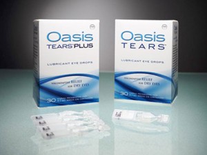
Not all eye drops are the same. Oasis TEARS is a new type of eye drop with a unique formula, optimized to provide long-lasting comfort. Some of the benefits of Oasis TEARS Lubricant Eye Drops:

  • Oasis TEARS offers instant relief of dry eye symptoms
  • It is natural and contains no damaging chemicals
  • Provides prolonged comfort with fewer applications
  • Provides moistening and lubrication to the surface of the eye
  • Eliminates the source of toxic irritation with preservative-free options

Oasis TEARS Lubricant Eye Drops coat, lubricate, and moisten delicate eye tissue. With each blink of your eye, a special substance in Oasis TEARS combines with the active ingredient, glycerin, to keep tears on the eye surface for a long time. This unique combination coats and recoats the surface of the eye for continued relief of dry and, irritated eyes. Use Oasis TEARS to relieve all dry eye symptoms any time of day. They work well for tired eye and discomfort related to computer vision syndrome.

Westside Optometry is pleased to offer Oasis TEARS for dry eye relief. To learn more about Oasis TEARS click here

What are Cataracts?

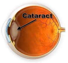
A cataract is a clouding of the lens in your eye that affects vision. Most cataracts are related to aging. To understand how a cataract can affect your vision, it helps to understand a little about the structure of your eye. The lens is clear and lies between the iris and the pupil. It works much like a camera lens, focusing light, or an image, onto the retina. The retina is the light-sensitive tissue at the back of the eye. Once the light or image reaches the retina, it is changed into nerve signals that are sent to the brain. Besides focusing light on the retina the lens also adjusts the eye’s focus, letting us see things clearly both up close and far away. The lens must be clear for the retina to receive a sharp image. The lens is made of mostly water and protein. The protein is arranged in a precise way that allows light to pass through it without distortion. But as we age, proteins in the lens clump together and darken, causing a cataract. If the lens is cloudy from a cataract, the image you see will be blurred and colors may appear faded.

How is the cataract removed?

A small incision is made on the side of the cornea, the clear, dome-shaped surface that covers the front of the eye. The surgeon inserts a tiny probe into the eye. This device emits ultrasound waves that soften and break up the lens so that it can be removed by suction. Most cataract surgery today is done by phacoemulsification, also called “small incision cataract surgery.”

After the natural lens has been removed, it often is replaced by an artificial lens, called an intraocular lens (IOL). An IOL is a clear, plastic lens that requires no care and becomes a permanent part of your eye. Light is focused clearly by the IOL into the retina, improving your vision. You will not feel or see the new lens. Depending on your vision and needs, you may be a candidate for a special type of IOL that either corrects astigmatism or acts like a bifocal.

Cataract removal is one of the most common operations performed in the United States. It is also one of the safest and most effective types of surgery. In about 90 percent of case, people who have cataract surgery have better vision afterward.

Twitching Eyelid

An eyelid twitch or tic is an involuntary spasm of the eyelid muscles. I’ve noticed twitches tend to occur in the same location. The most common causes are fatigue, stress, caffeine and eyestrain. So the best treatments include prevention of the causes: relax, get more sleep and consume less caffeine.

There are more serious twitches that involve the face and cause the eye to close with each spasm. Neurological causes must be ruled out. Temporary treatment is an injection with botulinum toxin (Botox). This relieves the spasm and allows the muscles to relax.

Most eyelid twitches are more annoying than serious.

Protecting Children’s Eyes from Sun Damage

Parents generally take precautions to protect their children’s skin from the sun; it is equally important to make every effort to protect children’s eyes from the sun’s harmful ultraviolet (UV) rays and glare. Just as we may not see the damage to our skin for years, we also may not see the damage to our eyes for some time. But each part of the eye can be damaged by chronic or high-dose exposure to UV radiation. Even the glare of an overcast day can add to the cumulative affect. Possible effects of long-term sun exposure to the eyes include:

  • The development of skin cancer on the eyelids
  • Changes to the surface of the eye, such as non-cancerous growths on the white of the eye
  •  Over time, the lens becomes more opaque and decreases vision forming cataracts
  • Damage to the retina, which can lead to age related macula degeneration, the leading cause of legal blindness in the US in people over 65 years.

In fact studies conducted over the last 30 years have proven that many of the serious eye diseases associated with aging are at least partially a result of years of sun exposure. Protecting children’s eyes are especially important due to two significant factors:

  1. Children spend more time outdoors. The average child receives 80% of his lifetime UV exposure before the age of 20.
  2. More damaging rays are transmitted through the young child’s crystalline lens to the retina. The eyes of children 10 years and younger transmit 75% of these rays; people 25 years or older only transmit 10%.

The simplest and most efficient way to prevent or delay the onset of   progression of various eye diseases is by using sunglasses or photochromic protective lenses designed to protect all damaging sun rays from reaching the eyes, ideally starting in early childhood. For a child who already wears corrective lenses, this can be achieved with polycarbonate photochromic lenses, which change color according to UV exposure. For a child who does not wear corrective glasses, a high quality pair of sunglasses is recommended.


High Blood Pressure and the Eyes

High Blood Pressure (HBP) is a serious condition that can lead to coronary heart disease, heart failure, stroke, kidney failure and vision loss. “Blood Pressure” is the force of blood pushing against the walls of the arteries as the heart pumps blood. If this pressure rises and stays high over time, it can damage the body in many ways.

About 1 in 3 adults in the United States has HBP. The condition itself usually has no signs or symptoms. You can have it for years without knowing it. During this time, the HBP can damage your heart, blood vessels, kidneys and eyes.

Blood Pressure tends to rise with age. Sometimes medication can control it. Following a healthy lifestyle helps people delay, prevent or control the BP.

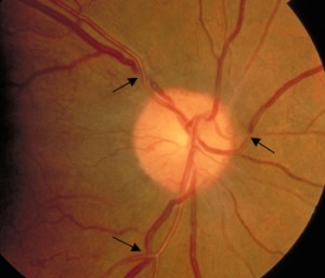
Hypertension can cause damage to the blood vessels in the retina. This picture of the retina shows an eye with hypertensive retinopathy. The arrows point to arteries and veins crossing each other. In a healthy eye the vessels run parallel. The crossings are dangerous because one of the vessels can be occluded. If an artery is blocked, blood will not flow to the retina beyond the occlusion and the retina will die. This causes a permanent blind spot. If a vein is occluded there will be bleeding which may cause a chain of negative events including loss of vision, abnormal vessel growth and glaucoma.

Hypertensive retinopathy typically won’t have any symptoms. It is found during a dilated eye exam. The earlier it is detected the sooner the blood pressure can be controlled preventing vision loss, stroke and death.

The best way to prevent hypertensive retinopathy is by keeping blood pressure controlled by diet, exercise and taking medications as prescribed.

Wearing Contact Lenses Overnight

Have you ever wondered what would happen if you wore your contact lenses overnight? Unexpectedly stayed the night at a friends house and didn’t have anywhere to put your lenses? Enjoying a  late night and fall asleep with your contacts on?

It happens, contact lenses get worn too long and sometimes over night. Some contact lenses are better for this than others. I prescribe and fit people with the intention of overnight use. I also warn patients about the risks they are taking with their eyes.

Depending on the contact lens type and material, oxygen is reduced to the cornea. Movement of the contact with each blink aids the transmission of oxygen. When you are sleeping with the contact lens, the eye is closed and no oxygen gets to the cornea. There is no blinking either so the lens doesn’t move. The reduction in oxygen causes the cornea to swell, if the lens allows plenty of oxygen to get to the cornea during the day, the cornea will deswell and return to normal. However, if the cornea remains swollen, and you sleep in the lens consecutive nights, blood vessels will begin to grow into the cornea. Sometimes the eye responds by becoming inflamed, red and painful.

Another risk of leaving the lenses in overnight is infection. Typically when a lens is removed it is thrown away or placed in disinfecting solution. In the morning a new lens or a clean, disinfected lens is applied to the cornea. Microbials build up on a lens that is worn continually. If your cornea is compromised by a scratch or edema, the bacteria will infect the eye.

If you have been prescribed extended wear contact lenses, stay compliant to your wear schedule, once in awhile overnight or 6 consecutive nights. Clean and replace your lenses as prescribed. If you are wearing your daily wear contacts overnight, let’s talk about a safer option. This may include a higher oxygen material or even LASIK. Sometimes the prescription can be a limiting factor. If the prescription is higher,  farsighted or is toric and corrects astigmatism, the lenses will be thicker and reduce oxygen transmissability.