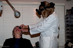
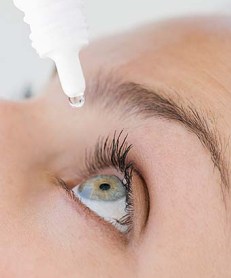
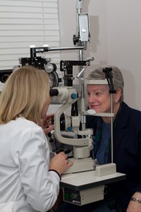
A comprehensive eye examination includes pupil dilation. Using eye drops, the pupil is temporarily and painlessly enlarged to increase the view into the eye. This gives me the opportunity to thoroughly examine the structures inside the eye: optic nerve, blood vessels, macula and retina. With the pupil dilated, I can get a 3-dimensional view and better evaluate the eye for changes or the potential for problems. The effect lasts up to 4 hours and will reduce the ability to focus on near objects. It will be helpful to bring sunglasses and make arrangements accordingly.
Category: Eye Health
Computer Vision
Computer Vision
Computer technology has evolved faster than the human eyeball. Our eyes accommodate our reading needs by focusing downward and approximately 16 inches away. Most computer monitors sit about 18 to 24 inches away from our eyes and only a few degrees below eye level. This is not much of a problem if your visual system is working accurately, but if you have a little refractive error such as astigmatism, farsightedness, or a difference between the two eyes, the result may be headaches, eyestrain and frustration.
If you are 40 years of age or older, the stress of viewing a computer screen is probably compounded by presbyopia: your eyes no longer accommodate, or focus, to read. (You may have noticed that your arms seem shorter.) Bifocals are often prescribed between the ages of 40 and 50, to allow you to see a distant object and also focus downward and close-up.
For someone who spends more than a couple of hours a day in front of a computer screen, a pair of glasses designed specifically for that task is the best solution. Do you wear cleats for golf? Flip-flops at the beach? Old tennis shoes to wash the dog? Do you have special shoes for special events? One pair of shoes can not fill every need; a single pair of glasses does not either.
A lens designed especially for the computer is recommended. It may be a single vision lens or a specifically designed multifocal.
A few more tips for comfortable computer use:
• Modify the room lighting to reduce glare and reflections.
• Anti-glare coatings can also eliminate reflections from windows, overhead lights and other computer monitors
• Place reference materials close to the screen so you do not have to turn your head back and forth to read.
• Use high-index materials to keep lenses thinner and lighter in weight.
• Take breaks to rest your eyes.
Visual Field Testing
Visual Field Testing
A perimeter is used to test your visual field. Your visual field includes both your central and side vision. During this test, while you are seated comfortably, you will be asked to look straight ahead at a central fixation target. You will be instructed to press a button when you become aware of a small shimmering light anywhere within your peripheral field. It’s important that you keep your eyes focused on the central target throughout this examination so that an accurate reading of your visual field is obtained.
This test can detect vision loss, even if the loss is far to the side or quite subtle. Even a large and profound vision loss in one eye’s visual field can go unnoticed by a patient because the visual field of the opposite eye overlaps it to a great extent. One eye simply “fill in” the area of vision loss present in the opposite eye. This is why the side-vision test is always administered to each eye separately.
The nerve pathways from the retina to the part of the brain where vision is interpreted are very uniform from person to person. Because of this uniformity, visual field analysis has a great deal of diagnostic value. Glaucomatous visual field loss usually follows a particular pattern. Other diagnostic patterns of visual field loss can indicate retinal detachment and neurological diseases, adding significantly to the value of this test. The nerve pathways for vision are long and cross over many brain structures on their route from the eye to the very back of the brain (visual cortex). Neurologists make great use of visual fields to help them determine where in the brain a tumor or other pathology is located.
Family History and Glaucoma
Family History and Glaucoma
 According to a study reported at the World Glaucoma Congress, the most efficient way to detect Primary Open-Angle Glaucoma (POAG), which is the most common form of glaucoma, is by knowing the medical history of close relatives with the disease. If you have a parent, sibling or child with POAG you have a greater than 25% chance of developing the disease. Others at risk include African Americans over 40, and anyone over 60, especially Mexican Americans. The earlier POAG is detected, the better the prognosis for retaining vision throughout one’s life. POAG occurs when the fluid that nourishes the eye cannot drain properly and the resulting increased pressure inside the eye damages the optic nerve. This is painless and not visually noticeable at first, but will lead to vision loss and blindness if left untreated. Increased pressure in the eye isn’t a sure sign of POAG, but it is a sign of increased risk for the disease.
Early diagnosis is of utmost importance. Dr. Griffith examines different parts of your eye to determine if you have or are at risk for developing POAG. Detection and diagnosis relies on tests for pressure within the eye (Tonometry), corneal thickness (Pachymetry) and observation of the optic nerve (Ophthalmoscopy) and visual fields, including peripheral vision. Because of the increased risk among family members, a review of family history is also part of the screening. (Likewise, a positive diagnosis of POAG would be valuable information for close family members.)
Vision lost to glaucoma cannot be restored. The goal with treatment is to slow the disease progress and prevent further vision loss. The most common treatment is a prescription eye drop or pill meant to reduce pressure in the eye. It is important for Dr. Griffith to be aware of other medications you are taking in order to fi nd a compatible treatment for the glaucoma; fortunately there are usually several options. Additional treatment includes laser surgery or sometimes more traditional surgery; both physically alter and improve the drainage structure in the eye. In conclusion, regular eye examinations are a certain age is vital to prevent unnecessary vision loss. If you are newly diagnosed with POAG or any form of glaucoma, Dr. Griffi th can give you much more detailed information.
Dry Eye Syndrome
 Dry Eye Syndrome
Red, gritty and scratchy eyes can have a number of causes and possible treatments. In addition to discomfort and fluctuating vision, dry eyes can lead to styes and infection. Regular eye examinations can prevent complications and provide the opportunity for you to get help with treatment.
If you have dry eyes, you may notice that the discomfort worsens as the day progresses. Air conditioning, smoke, drafts, and cold temperatures become difficult to tolerate. It may feel like there’s a foreign body in your eye, or your eyes may have a sandy feeling. In spite of the fact that you are suffering from “dry” eyes, you may find that your eyes are watering reflexively and that strands of mucous are accumulating.
What’s Going On? Normal tear film consists of three layers: mucin, aqueous, and lipid. Abnormalities in production, content, or distribution of these three layers or in eyelid function will cause the various conditions commonly known as dry eye.
- Lipid Problems: These are the most common cause of dry eye. Glands in the eyelid produce lipids. Greasy lotions, incomplete removal of makeup, or skin conditions like dandruff can plug the glands and prevent lipids from secreting.
- Aqueous Deficiency: This can be a side effect of certain medications you’re taking. The culprits include antihistamines, diuretics, hormones, and psychotropics.
- Mucin Deficiency: Chronic infection or trauma to the eye can cause a lack of mucin. Autoimmune diseases negatively effect the mucin layer too.
- Eyelid Problems: The eyelid may turn in or out as a result of the aging process or of scarring. Tears then spill over the eyelid, allowing the eye to become dry. Depending on the severity of the situation, this problem can be treated with plastic surgery.
Also, dry eye can be aggravated by inflammation of the eyelid margin. In this case, the lid, where the eyelashes attach, will be red and irregular.
No one treatment for dry eye has proven completely successful. A combination of the following may improve comfort: good eyelid hygiene, the use of artificial tears and lubricating ointments, newer agents designed to heal tissue such as Cyclosporine, supplements and punctal occlusion (the insertion of a silicone plug into the tiny opening that drains the tears). Examining controllable factors such as medications, topical creams and lotions, and environment enhances treatment success.
Allergies

Your eyes can develop allergic reactions just like your body does. You may experience symptoms such as itching, burning and watering eyes. Here is a look at some of the most common ocular allergies and their symptoms.
Your Eyes and Allergies
Hay Fever or Dust Conjunctivitis
Conjunctivitis is an inflammation of the conjunctiva, the thin, transparent mucous membrane draped over the eye. Conjunctivitis is commonly called “pink eye.” Plant pollen, house dust and animal dander may trigger a reaction in sensitive eyes and people. The reaction usually starts a short time after your exposure, and may include tearing, itching and swelling. Your symptoms may be worse at the end of the day.
Giant Papillary Conjunctivitis
Contact lens wearers know this allergy more commonly as GPC. Among the things that may cause GPC are improper cleaning of contact lenses, infrequent contact lens replacement, and wearing contact lenses for too many hours. You have a greater risk of this allergy if you have asthma, hay fever or animal allergies. GPC may occur months or even years after you begin contact lens wear. GPC’s symptoms are itchy eyes after removing your lenses, mucus discharge in the morning, sensitivity to light and uncomfortable lenses. Your vision may blur due to deposits on the lenses or lens movement caused by tiny allergic bumps (papilla) on the inside of your upper eyelid.
Contact Dermatitis
Allergic contact dermatitis occurs when you come into contact with something that irritates your skin. Your eyelids and conjunctiva are very sensitive. Signs of this reaction include swelling, redness, scaly skin and blistering. Symptoms are itching and pain. The reaction usually develops about 12 to 72 hours after you are exposed.
Recommendations to Reduce Allergic Reactions
- Avoid exposure to allergen
- Rinse eyes with sterile saline solution
- Cold compresses – a couple of ice cubes in a clean washcloth
- Maintain contact lens integrity by replacing and cleaning the lenses as prescribe
- Reduce irritation with proper eyelid and eyelash hygiene
- Control ocular dryness with artificial tear drops, proper hydration and supplement
- Begin prescription allergy drops at the first signs of a reaction
Dr. Griffith can help you manage your ocular allergies.

