I occasionally have patients ask if their cataract is “ripe.” There is some misunderstanding, cataracts don’t ripen. The crystalline lens does change and causes vision problems. To read more about what a cataract is click here.
Cataracts can be removed whenever the patient and surgeon agree that they should be removed. Generally, the best time to remove the cloudy crystalline lens (AKA cataract) is when the vision is compromising your quality of life. This includes the inability to drive at night due to excessive glare. Other causes of blurry vision must be explored before cataract surgery. Macular degeneration can reduce the quality of vision, especially for reading. Removing cataracts will not improve the clarity in this case.
Category: Eye Examination
Maintaining Clear Vision
Age-related eye diseases such as macular degeneration and cataracts commonly cause impaired vision and blindness in older adults. But lifestyle changes, including good nutrition, could help delay or prevent certain eye problems.
Besides adopting a healthy diet, you also can protect your eyes by avoiding intense ultraviolet (UV) light, quitting smoking and getting regular checkups that may help detect chronic diseases contributing to eye problems. Diabetes, for example, increases your risk for age-related eye diseases and may cause diabetic retinopathy.
Regular eye exams, too, are essential for maintaining eye health as you grow older. If eye problems and chronic diseases are detected early enough, appropriate treatment may prevent permanent vision loss.
What is Glaucoma?
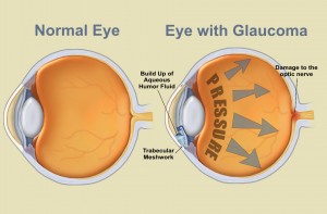
January is glaucoma awareness month. Glaucoma is often called “the sneak thief of sight.” 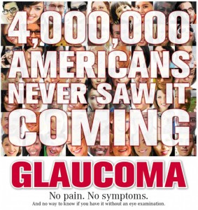 That’s because people usually do not notice any signs of the disease until they have already lost significant vision. Once lost, vision can’t be restored. More than 2.7 million Americans age 40 and older have open angle glaucoma, the most common form of glaucoma. At least half don’t even know they have it.
Glaucoma is an eye disease that causes loss of sight by damaging a part of the eye called the optic nerve. This nerve sends information from your eyes to your brain. When glaucoma damages your optic nerve, you begin to lose patches of vision, usually side vision (peripheral vision). Over time, glaucoma may also damage the central vision. You may not notice a loss of side vision until you have lost a great deal of your sight. When checking for glaucoma, I measure the intra-ocular pressure, dilate the pupil to look for damage to the optic nerve and test peripheral vision. To learn more about how I identify glaucoma and how it is treated, click here
Who is at risk for glaucoma?
Below are the risk factors for glaucoma:
Age – The older you are, the greater you are at risk (especially if you are over 60 years old). African Americans are at a greater risk at a younger age starting at age 40 and older.
Race – African Americans age 40 and over are 4-5 times more likely to have glaucoma than others. Hispanics are also at increased risk for glaucoma as they age. Those of Asian and Native American descent are at increased risk for angle closure glaucoma.
Family history – If you have a parent, brother or sister with glaucoma, you are more likely to get glaucoma. If you have glaucoma, inform your family members to get complete eye exams.
Medical history – You are at risk if you have a history of high pressure in your eyes, previous eye injury, long term steroid use, or nearsightedness.
There are no symptoms for glaucoma until irreversible vision loss occurs. Have your eyes check.
New Year’s Resolutions for Better Vision

2013 can be the year you have the best vision possible. Below are some suggestions to help you.
Improve Your Eye Health
- Eat smart. Diet and nutritional supplements go a long way in promoting eye health. Studies show a diet rich in fruits, leafy vegetables and omega-3 fatty acids may reduce your risk of eye problems like macular degeneration and dry eye syndrome.
- Get moving. Research has shown higher levels of physical exercise can reduce certain risk factors for glaucoma, as well as macular degeneration.
- Quit smoking. Put simply, smoking harms your vision. Studies show smoking dramatically increases the likelihood of developing cataracts, macular degeneration,uveitis and diabetic retinopathy.
- Protect your eyes from the sun (and make sure your kids do, too). Always wear sunglasses with UV protection when outdoors — no matter what time of year — to shield your eyes from UV rays. This may reduce your risk for cataracts and macular degeneration.
- Schedule an eye exam for everyone in your family. Kids and seniors, especially, should have comprehensive annual eye exams to monitor vision changes. Also, have your family doctor screen you for diabetes and hypertension — if left untreated, these diseases can lead to serious eye problems.
- Start using safety eyewear for lawn-mowing, home repairs and other chores. Experts say 90 percent of eye injuries requiring a visit to the emergency room can be prevented with proper safety eyewear.
- Properly care for your contact lenses. Dirty contact lenses, even if they are not uncomfortable, can cause serious eye infections. Clean your contact lenses and contact lens case properly, and always replace your contacts as recommended.
- Reduce computer eye strain. Rest your eyes from computer work every 20 minutes to relieve computer vision syndrome and avoid dry, red eyes. Also, ask your eye doctor about stress-relieving computer glasses.
- If you’ve been putting up with contact lens discomfort, dry eyes, eye allergies or blurry vision, talk to me about changes you can make to improve or eliminate these problems.
Improve Your Vision
- Blurriness? If your contacts or glasses are no longer doing their job, you may need a new
 prescription and/or a different lens design.
 - Upgrade your contact lenses. Contact lenses come in a wide variety of materials, replacement schedules and wearing times — not to mention the array of color contact lenses and special effect contacts available.
- With the advancement in contact lens technology, there’s sure to be a type of contact lens that suits your individual requirements and lifestyle.
- Try eyeglass lens coatings. Various lens coatings keep your field of view clear by reducing reflections, fogging and scratches. And eliminate glare during outdoor activities with polarized sunglasses.

- Consider sports-specific eyewear. For athletes and sporting enthusiasts, there are performance-enhancing frames and lenses designed specifically for different sports and outdoor activities.
- Make sure your sports eyewear includes lightweight, impact-resistant polycarbonate or Trivex lenses for comfort and safety.
Improve Your Appearance
- Upgrade your eyewear. Get with the times and refresh your look, as well
 as take advantage of the latest in lens and frame technologies.
 - If you have a strong prescription, try high index eyeglass lenses. High index lenses provide the same optical power as regular ones, but are thinner and lighter.
- Considering LASIK? If you’re tired of wearing glasses or contacts, ask your us if you are a good candidate for LASIK or other vision correction surgery.
Happy New Year
Westside Optometry Hours through the Holidays
Monday, December 31st 8:30 – 12:00
Tuesday Closed for New Years Day
Regular hours resume
Wednesday, January 2nd 8:30 – 5:00
Thursday, January 3rd, 9:00 – 6:00
Friday, January 4th, 8:30 – 12:00
Saturday, January 5th, 8:00 – 12:00
We wish you a very Happy New Year
Color Vision Deficiency
Color vision deficiency is often called “color blindness” by mistake. Color vision deficiency describes a number of different problems people have with color vision. Abnormal color vision may vary from not being able to tell certain colors apart to not being able to identify any color.
Color vision deficiency affects an estimated 8% of males and fewer than 1% of females. Most color vision problems run in families and are inherited and present at birth.
A child inherits a color vision deficiency by receiving a faulty color vision gene from a parent. Abnormal color vision is found in a recessive gene on the X chromosome, therefore, color vision deficiency is a sex-linked condition. Because males only have one X chromosome, they are more likely to have color vision deficiencies than females.
Heredity does not cause all color vision problems. One common problem happens from the normal aging of the eye’s lens. The lens is clear at birth, but the aging process causes it to darken and yellow. Older adults may have problems identifying certain dark colors, particularly blues. Certain medications as well as inherited or acquired retinal and optic nerve disease may affect normal color vision.
At Westside Optometry, we test our young boys and some girls for color vision deficiency. Any child who is having difficulty in school should be checked for possible visual problems including color vision impairment. If there is a family history of color deficiency or your child is having problems identifying colors, let us know so he/she can be tested.
The specialized cells in the retina are called rods and cones. You use these cells for normal vision. Rods are useful for night vision and working in dim light. Cones are responsible for color vision. They work best in daylight. Three types of cone pigments are present in normal vision. These are sensitive to either blue, green or red colored objects. Together, they let you see a wide range of colors, from purple through red.
For normal color vision, all three cone pigments must work correctly. When a cone pigment is abnormal or missing, a type of color vision deficiency results. For example, the most common deficiency causes confusion between red and green colors. In rare cases, a person is born without any cones. These people are truly “color blind.” They see the world in shades of gray and suffer significant visual impairment. Most types of color vision deficiency are present at birth. There are also some types caused by aging, eye disease or injury.
Color Vision Testing
There are several ways to test color vision. Simpler tests involve colored  figures (either shapes or numbers) placed against a busy, patterned background. A person with normal color vision can see the figures against the background. Those with color vision deficiencies cannot see the symbols.
At Westside Optometry we do a more specific test that requires placing colored disks in order, from one shade to another. People with different color deficiencies place the disks in varying order.
Unfortunately, there is no cure for hereditary color vision deficiency. Many people with color vision deficiency develop their own “system” or learn to  identify colors by other means. Some people learn to tell colors apart by brightness and location. A stoplight, for example, is designed so the colors are always in the same order.
Diabetic Retinopathy Continued
This is a continuation of my last blog post about diabetic retinopathy. I want to stress that diabetic retinopathy is the number 1 cause of new cases of blindness for adults 20-70 years old. The increasing number of people with diabetes means the number of people who will develop diabetic retinopathy will also increase. This is significant because severe vision loss can be prevented 90% of the time.
It is my job as an optometrist to identify and detect diabetic retinopathy. When I see diabetic retinopathy, I have to decide when to refer for further evaluation and/or treatment.
PREVENTING VISION LOSS
The most important thing you can do to prevent vision loss from diabetes is have a dilated eye examination every year.
If you notice changes in your vision or it seems blurry, call your eye doctor immediately. To read more about preventing eye complications from diabetes click here.
TREATMENT
If I think treatment is indicated, I will refer you to a retinologist. A retinologist is an ophthalmologist, who treats conditions of the vitreous and retina, both effected by diabetic retinopathy. He or she will chose the best treatment option.
A laser may be used to stop blood vessels from leaking. It may also be used over a larger part of the retina to reduce the growth of abnormal blood vessels. Laser maintains sight, but the side effects can include, blind spots in the vision and reduced vision.
Corticosteroid injections into the eye provide a temporary treatment. To maintain control of the retinopathy, repeated injections are necessary every 6-8 weeks. The continued use of corticosteroids increases the risk of developing cataracts and glaucoma.
Another treatment is the injection of anti-vascular endothelial growth factor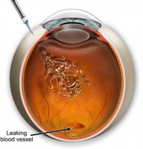 (anti-VEGF) to prevent the blood vessels from leaking. This treatment also needs to be repeated every 6-8 weeks.
The retinologist will often use a combination of the above treatments to yield the best results.
Researchers continue to look for therapies with long-term results and minimal side effects.
Diabetic Retinopathy
Westside Optometry continues to recognize November as Diabetes Awareness Month, although diabetes is something that deserves attention all 365 days of the year. If you have diabetes, I will dilate your eyes at least once a year. Diabetes can affect many organs of the body, in the eyes it causes blindness.
Many problems develop in the retina due to diabetes. The possibilities include abnormal blood vessel growth, hemorrhages and lipid leakage. If these problems are allowed to continue without treatment they will cause scarring which leads to detachment of the retina. Another complication is the leakage of fluid under the macula which will severely reduce vision.
Not all these conditions will have symptoms. Only when the bleeding or fluids reach a certain size will you notice blur or dark spots. The earlier changes in the retina are detected, the better treatment results will be.
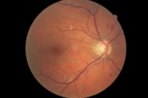
The picture above shows some of the changes diabetes causes in the retina. There is bleeding and some areas where blood isn’t flowing properly (ischemia). This patient did not notice any changes in his vision. If you have diabetes, don’t wait until you have vision changes, it may be too late.
How to Prevent Eye Complications due to Diabetes
There are steps you can take to avoid eye problems due to diabetes.
First and most important, keep your blood sugar levels under tight control. In the Diabetes Control and Complications Trial, people on standard diabetes treatment got retinopathy four times as often as people who kept their blood sugar levels close to normal. In people who already have retinopathy, the condition progressed in the tight-control group only half as often.
These impressive results show that you have a lot of control over what happens to your eyes. Also, high blood sugar levels may make your vision temporarily blurry.
Second, keep blood pressure under control. High blood pressure can make eye problems worse.
Third, quit smoking.
Fourth, see your optometrist at least once a year for a dilated eye exam. 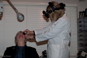 Having your regular doctor look at your eyes is not enough.
Fifth, see your optometrist if:
- your vision becomes blurry
- you have trouble reading signs or books
- you see double
- one or both of your eyes hurt
- your eyes get red and stay that way
- you feel pressure in your eye
- you see spots or floaters
- straight lines do not look straight
- you can’t see things to the side as you used to
Don’t procrastinate. If you have diabetes and haven’t had a dilated eye exam in the last 12 months, schedule an eye exam now.
For more information, check out the American Diabetes Association.
High Blood Pressure and the Eyes
High Blood Pressure (HBP) is a serious condition that can lead to coronary heart disease, heart failure, stroke, kidney failure and vision loss. “Blood Pressure” is the force of blood pushing against the walls of the arteries as the heart pumps blood. If this pressure rises and stays high over time, it can damage the body in many ways.
About 1 in 3 adults in the United States has HBP. The condition itself usually has no signs or symptoms. You can have it for years without knowing it. During this time, the HBP can damage your heart, blood vessels, kidneys and eyes.
Blood Pressure tends to rise with age. Sometimes medication can control it. Following a healthy lifestyle helps people delay, prevent or control the BP.
Hypertension can cause damage to the blood vessels in the retina. This picture of the retina shows an eye with hypertensive retinopathy. The arrows point to arteries and veins crossing each other. In a healthy eye the vessels run 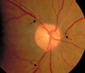 parallel. The crossings are dangerous because one of the vessels can be occluded. If an artery is blocked, blood will not flow to the retina beyond the occlusion and the retina will die. This causes a permanent blind spot. If a vein is occluded there will be bleeding which may cause a chain of negative events including loss of vision, abnormal vessel growth and glaucoma.
Hypertensive retinopathy typically won’t have any symptoms. It is found during a dilated eye exam. The earlier it is detected the sooner the blood pressure can be controlled preventing vision loss, stroke and death.
The best way to prevent hypertensive retinopathy is by keeping blood pressure controlled by diet, exercise and taking medications as prescribed.

