Flashes and Floaters

Floaters are spots which can move about in the vision and which are sometimes accompanied by flashes of light. To understand these symptoms it is necessary to understand the basic anatomy of the eye.

Anatomy of the Eye:
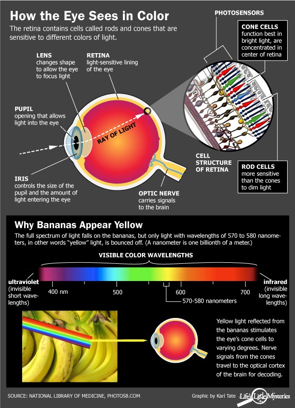
The structure of the eye resembles that of a hollow ball. The Parts of the Eye
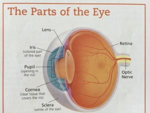
central hollow is filled with a clear jelly called the vitreous. In the front part of this ball there is an opening called the pupil leading to the outside. Looking the inside of the hollow is a thin layer called the retina which is the part of the eye with which a person sees. When we are children the vitreous is solid like cold jello.

Floater Formation:

As part of normal changes, the vitreous gel becomes liquefied. This does not occur all at one time so there are pockets of liquefied vitreous next to solid vitreous gel with a filament-like membrane between. When the eye moves the liquid moves easily and sloshes around, causing the filament-like membranes to move and the movement of the membrane casts a moving shadow on the retina which we see as a floater. Where the membranes are attached to the retinal surface, the movement causes a tugging, pulling or traction on the retina. When the retina is pulled or bumped we see the response as a flash of light. Therefore, when the eye moves we see floaters and sometimes we also see light flashes.

If the membrane attached to the retina pulls strongly enough, it can produce a tear or hole in the retina. When a tear forms, there may be a small amount of bleeding, and blood particles moving around in the liquid create floaters which are like a shower of tiny black spots.

Significance of Floaters:

Floaters are not by themselves dangerous, but are a warning that a hole in the retina might exist or that other changes might have taken place. Because of this, it is important for anyone who has recently developed floaters to have a dilated eye examination, including examination of the far edges of the retina where tears are most likely to occur. In the majority of cases this examination will not reveal any holes. If, however, a hole is found in the retina, it usually can be treated with either laser before a more serious problem, such as a retinal detachment, develops.

Light Flashes:

These also can be associated with shrinkage of the gel-like substance (vitreous) as it moves and bumps the retina. Flashes can be caused by rubbing the eyes too hard. Retinal hole formation can also cause light flashes, and so the presence of this symptom requires a careful retinal examination. Flashes can occur from other causes as well.

Follow-up of Floaters and Flashes:

Repeat dilated eye examinations are indicated if suspicious signs are found during an eye examination or if later the patient notices either a sudden increase in floaters and flashes or a veil falling into the vision.

If you are experiencing either new floaters and/or flashes of light, call to have a dilated eye examination sooner than later. Early treatment of holes and retinal tears is less risky and more successful than treatment of a retinal detachment.

Keep Your Vision Healthy

Clear View

Did you know your eyes are windows to your general health? Many systemic conditions can be detected with a dilated eye examination. Looking into a dilated eye, I can see a view of the blood vessels and assess vascular health. Your eyes can tell a lot about your visual health and overall wellness.
Besides helping you see better, annual eye exams can aid in detection of serious eye conditions, like glaucoma and cataracts and health conditions like diabetes and high blood pressure. This is important since you won’t always notice the symptoms yourself – and some of these diseases cause irreversible damage.

If it’s been at least a year since your last eye examination, call the office and schedule an appointment. (707)762-8643. Schedule online.

What is Macular Degeneration?

Macular degeneration is a retinal disease which can lead to legal blindness. More specifically, it affects the macula, the portion of the retina important for detailed vision like reading.

There are two forms of macular degeneration: wet and dry. Dry macular degeneration is usually slower in destruction of the macula, causing gradual to little loss of central vision. The wet form is more aggressive and destroys the central vision faster. The wet form is associated with the presence of abnormal blood vessels growing somewhere within the layers of the retina. These abnormal blood vessels can leak and bleed.

ARMD wet before treatment

Dry macular degeneration causes a slower loss of tissue. While there is treatment for the wet form, there is no approved treatment for the dry form.

Drusen (800x533)

Symptoms of Macular Degeneration include blurry central vision and/or distortion. In some cases, there may be blind spots within the central vision. The vision loss in both types is progressive. It is the rate at which vision loss develops which differentiates the wet form from the dry. Remember, wet macular degeneration causes faster and more devastating loss of vision compared to the dry form.

Treatment for Macular Degeneration

There is no treatment for dry macular degeneration. While AREDS (Age-Related Eye Disease Study) vitamins may be indicated for a small subset of macular degeneration patients, it does not improve the condition or vision. The supplements are primarily preventative. There are different formulations of “eye vitamins,” but the contents of the supplement used for the study were 500 milligrams of vitamin C; 400 International Units of vitamin E; 15 milligrams of beta-carotene (often labeled as equivalent to 25,000 International Units of vitamin A); 80 milligrams of zinc as zinc oxide; and two milligrams of copper as cupric oxide. Copper was added to the AREDS formulations containing zinc to prevent copper deficiency anemia, a condition associated with high levels of zinc intake.

Other preventative measures for both types of macular degeneration include not smoking, regular exercise and a healthy diet.

The mainstay of treatment for wet macular degeneration involves injections into the eye with anti-VEGF (Vascular Endothelial Growth Factor) medications.

These medications (Lucentis, Avastin, Eylea) attach to the abnormal blood vessels, preventing additional leakage and bleeding. Often there can be improvement in vision.

Early treatment is key to maintaining vision. Regular dilated eye examinations are advised.

How Does Diabetes Damage Your Eyes?

November is Diabetes Awareness month. High amounts of blood sugar can harm the internal structures of the eye. Below are some of the things that diabetes can do to the eyes.

  • Blurry or Double Vision. Fluctuating blood sugar and fluctuating vision are connected. A change in glucose levels affects the eye’s ability to maintain sharp focus. It may take several months after your blood sugar is well controlled for your vision to stabilize.


  • Diabetic Retinopathy. The leading cause of diabetes-related vision loss is diabetic Diabetic Retinopathy retinopathy. This condition damages the network of blood vessels supplying the retina with blood and oxygen. It also causes abnormal growth of blood vessels on the retina. When these vessels leak they damage  the retina causing permanent vision loss.
  • (the picture on the left is an example of fluid and blood leakage in diabetic retinopathy.)


  • Cataracts. People with diabetes have a much greater risk of developing cataracts and at an earlier age. A cataract is clouding of the eye’s natural lens and results in glare and obscured vision.
  • Glaucoma. Diabetes also increases your risk of developing glaucoma. This condition results from an increase in fluid pressure inside the eyeball, which causes damage to the retina and optic nerve. If untreated, glaucoma leads to gradual but permanent vision loss.

Actions to take right now include the following: Eating sensibly, the American Diabetes Association recommends filling half your plate with vegetables, a quarter of the plate should be lean protein and the rest a carbohydrate. Make time for 30 minutes of exercise everyday. Take your medicine as prescribed and know your blood sugar level.

Preventing diabetes is the best way to avoid diabetic damage to the eyes, but if you already have diabetes, regular eye examinations can prevent permanent damage to the eye due to elevated blood sugar.


Eyes on the Road

Shorter days and rainy weather, both contribute to difficulty driving. A driver’s vision guides 90% of driving decisions, so good eyesight is essential for road safety.

On the Road

Start with an eye examination to assess clarity, peripheral vision and eye disease. Having the proper vision skills and prescription are important for many reasons:

Distance vision is imperative for judging distances and reaction time when driving. Poor distance vision increases the risk of making unreliable judgments with potentially disastrous results – even the simplest reactions take 0.4 seconds, so if distance vision is poor, signs or objects may not be seen until it is too late to react safely.

Good Peripheral Vision allows drivers to survey the area without looking away from the road. This includes early recognition of cross-traffic, pedestrians, animals, signs and changes in traffic flow. Impaired peripheral vision can result in the driver failing to react to a hazard on the far left or right, failing to stop at a stoplight suspended over an intersection, weaving while negotiating a curve, or driving too close to parked cars.

Safe Night Driving requires the ability to see in low light conditions and with poor contrast. Impaired night vision can result in tailgating, failing to steer when necessary (since it is difficult to see low contrast features such as edges or irregularities in the road surface), and recover from glare of oncoming headlights.

Glare refers to the disruption of vision due to veiling luminance. Important visual skills for drivers are glare resistance – the extent to which the driver can still see critical objects while facing a steady source of glare (setting sun, headlights), and glare recovery – the rapidity with which the driver’s functioning vision returns to what it was before the glare was encountered. If these skills are not adequate, the driver can miss curves, strike an animal or pedestrian or crash into slow moving or stopped vehicles.

Besides regular eye exams, additional tips for good vision while driving include:

  • Always wear sunglasses when driving to reduce glare
  • Keep headlights, tail lights , windshields and eye glasses clean
  • Keep side and rear view mirrors adjusted correctly to increase your field of vision and reduce glare from headlights
  • Always wear glasses or contact lenses if they have been prescribed for driving

20/20 Vision

During an eye examination, I always use the eyechart to measure the distance vision, usually with the most current glasses or contact lenses. This gives me a place to begin and tells me what sort of vision a person has and a quantity to compare. snellen chart
The classic example of an eye chart is the Snellen eye chart, developed by Dutch eye doctor Hermann Snellen in the 1860’s. There are many variations of the Snellen eye chart, but in general it has 11 rows of capital letters. The top row contains one letter (usually an E). the other rows contain letters that are progressively smaller.

In the United States, the standard placement of the eye chart is on a wall that’s 20 feet away from your eyes. Since most of our exam rooms are shorter than 20 feet we use mirrors to simulated the 20 foot distance. 20/20 vision is considered “normal vision”, meaning you can read at 20 feet a letter that most human beings should be able to read at 20 feet.

In the US a person is considered “legally blind” if his best-corrected visual acuity with glasses or contact lenses is 20/200 or worse. To get a driver’s license in California a person must have at least 20/40 best corrected acuity.

An eye chart is a measure of visual acuity only. It is an aid to prescribing glasses and contact lenses. But eye charts don’t measure peripheral vision, depth perception, or color perception. They also don’t determine the ocular health.

The visual acuity measurements done with the Snellen eyechart are simply a starting place for any eye examination, a method to quantify it.

Good Vision and Learning

Vision is a dominant process in the growth, development and daily performance of children. Many children with undetected vision problems struggle in the classroom. Some symptoms of learning related vision problems are:

  • Trouble finishing written assignments
  • Losing ones place when reading
  • Having a short attention span when doing close work
  • Skipping words when reading
  • Making errors when copying from a classroom board
  • Having greater potential than grades may indicate (under-performing)

Vision problems can affect comprehension performance in reading and manifest as social, eye-hand coordination, discipline, or emotional problems. From there, such vision problems can impact the rest of your child’s life and ability to succeed.


[heading centered=”yes” margin_bottom=”no”]What is Good Vision?[/heading]

Back to school Visual Acuity: It’s important to realize that good vision is more than 20/20 eyesight, Invented in the 1860’s, the term 20/20 indicates if you can see letters 3/8″ high at 20 feet. This does not take into account the eyes’ ability to see books or view the computer screen.

Eye Health: Eye disease can impair vision or lead to vision loss if not diagnosed and treated.

Visual Integration: The ability to process and integrate visual information, which includes and coordinates input from our other senses and previous experiences so that we can understand what we see.

Eye Teaming: The ability of the eyes to work properly together.

Eye Focusing: The ability of the eyes to focus and shift focus to near and distant points easily and effortlessly.

Eye Motility or Tracking: The ability of the eyes to move together across a page of print, to directly view an object, to move from one viewing area to another, or follow a ball.

The good news if that with early diagnosis and appropriate, comprehensive intervention, the prognosis is good in a majority of cases. Schedule your child’s eye examination to make the most of the new school year.

Contact Lens Prescriptions

Contact lens prescriptions generally expire on a yearly basis, unless otherwise specified. Seeing your eye doctor regularly for a comprehensive eye exam will not only keep your prescription updated, and evaluate your ocular health, but an eye exam will also help identify and lead to diagnosis of other health concerns such as hypertension and diabetes. At your eye exam Dr. Griffith may recommend a newer/better contact lens option for you, too.
A contact lens is a medical device and can be worn to correct vision as well as for cosmetic or therapeutic reasons. In the United States, all contact lenses, even purely cosmetic ones, require a prescription. They must be properly fitted and prescribed by an eye doctor. soft CL

An eye examination is needed to determine an individual’s suitability for contact lenses. This typically includes a refraction to determine the proper power of the lens and an assessment of the health of the eye. Dr. Griffith will also ask questions about your lifestyle, general health and contact lens wearing goal. If you haven’t worn contact lenses before, training for application and removal of the contact lens is necessary. If the lenses are to be re-used, a care and disinfecting system is required too. A follow-up appointment will determine the proper fit and lens compatibility for your eyes. Contact lenses are not a “one size fits all” device. There are many parameters to each lens. Besides the power to correct the vision, material and edge design will effect the comfort. The size: diameter and curvature are factors in the fit and ultimately the response of the cornea and eyelids to the contact lens.

Prescriptions for contact lenses and glasses may be similar, but are not interchangeable.

How the Eye Sees Color

Color VisionThe 6 to 7 million cones in the human retina are responsible for color vision. The cones are photoreceptors concentrated in the central zone of the retina called the macula. The center of the macula is called the fovea, and this tiny (0.3mm diameter) area contains the highest concentration of cones in the retina and is responsible for our most acute color vision.

Inherited forms of color blindness often are related to deficiencies in certain types of cones or outright absence of these cones. Color blindness is not a form of blindness at all, but a deficiency in the way we see color. With this vision problem, individuals have difficulty distinguishing certain colors, such as blue and yellow or red and green.

Color vision deficiency is an inherited condition that affects males more frequently than females. An estimated 8 percent of males and less than 1 percent of females have color vision problems.

Red-green color deficiency is the most common form of color deficiency. Red-green color blindness is caused by a common X-linked recessive gene. Your mother must be a carrier of the gene or be color deficient herself. Fathers with this inherited form of red-green color blindness pass the X-linked gene to their daughters but not their sons, because a son cannot receive X-linked genetic material from his father.

Any time a mother passes along this X-linked trait to her son, he will inherit the color vision deficiency and have trouble distinguishing reds and greens.

There is no cure for color blindness. But some coping strategies may help an individual function better in a color-oriented world. Most people are able to adapt to color vision deficiencies without too much trouble. But some professions should be avoided, such as graphic designer, ship pilots, interior decorator and other occupations that  require precise differentiation of colors and depend on accurate color perception.

If the color deficiency is identified early enough in life, the individual may be able to compensate by training for one of the many careers that are not as dependent on the ability to see in a full range of colors.

It is possible to develop color vision problems later in life. Sudden or gradual loss of color vision can indicate any number of underlying health problems, such as cataracts. Color blindness can occur when changes due to aging  damage retinal cells. An injury or damage to areas of the brain where vision processing takes place also can cause color vision changes. If you feel your perception of color is changing, schedule an eye exam.

At Westside Optometry we use a desaturated color vision test that takes a couple of minutes.

Heart Disease and the Eyes

heart in hands

The eyes are the window to the soul

In honor of Valentine’s Day, I want to explain why the eyes are the window to the heart.
Abnormalities of the blood vessels in the retina can be used to indicate complications for diabetes, hypertension, stroke and heart disease.
The retina is a membrane that lines the eyeball and receives light and converts it into signals that reach the brain and result in vision. During a dilated eye exam, Drs. Griffith and Staton examine the retina and blood vessels. Changes in the vessels such as narrowing or ballooning is suspicious of cardiovascular disease. Systemic diseases, that affect the body or its organs, such as hypertension, diabetes, AIDS, Graves’ disease, lupus, atherosclerosis, multiple sclerosis, rheumatoid arthritis and sickle cell anemia often cause changes in the eye that show up as red dots or small blood clots. Blood vessels of the eyes are so predicative because they are part of the brain’s vascular system, so they share anatomical features and respond similarly to stress and disease.

Normal Retina Photo
Normal Retina

In fact, eyes are so transparent compared to the rest of the body that they are the only organ that allows doctors to directly see blood vessels. The use of digital fundus photography allows the doctor to analyze the retina and vessels and compare the pictures to previous photos taken.

Let us gaze into your eyes.